984
Views
70
CrossRef citations to date
0
Altmetric
Research Paper

BMP-2 inhibits the tumorigenicity of cancer stem cells in human osteosarcoma OS99-1 cell line

, , , , , & show all
Pages 457-463 | Received 03 Aug 2010, Accepted 03 Dec 2010, Published online: 01 Mar 2011

Figures & data

Figure 1 Expression of BMP receptors in human osteosarcoma tumorigenic ALDHbr cells. (A) Messenger RNA transcripts for BMP receptors. Lane 1: negative control; Lane 2: freshly sorted ALDHbr cells; Lane 3: MCF7 cells (positive control). Representative immunofluorescent staining of BMPR1A (B), BMPR1B (C) and BMPR2 (D) in freshly sorted ALDHbr cells. Representative immunofluorescent staining of BMPR1A (E), BMPR1B (F) and BMPR2 (G) in unsorted cells from OS99-1 xenografted tumors, and there was no difference on the expressions of those three BMP receptors between the ALDHbr cells and the unsorted cells. The nuclei counterstained with 4′,6-diamidino-2 phenylindole (DAPI) (blue areas).
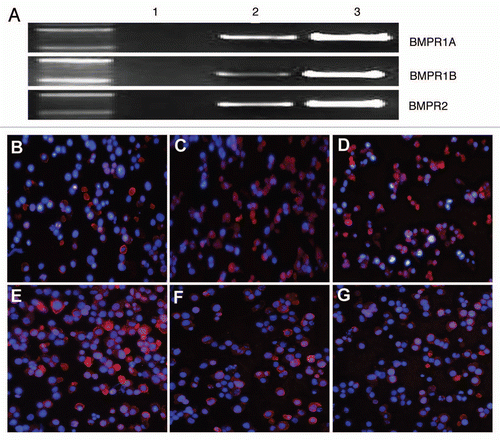
Figure 1 Expression of BMP receptors in human osteosarcoma tumorigenic ALDHbr cells. (A) Messenger RNA transcripts for BMP receptors. Lane 1: negative control; Lane 2: freshly sorted ALDHbr cells; Lane 3: MCF7 cells (positive control). Representative immunofluorescent staining of BMPR1A (B), BMPR1B (C) and BMPR2 (D) in freshly sorted ALDHbr cells. Representative immunofluorescent staining of BMPR1A (E), BMPR1B (F) and BMPR2 (G) in unsorted cells from OS99-1 xenografted tumors, and there was no difference on the expressions of those three BMP receptors between the ALDHbr cells and the unsorted cells. The nuclei counterstained with 4′,6-diamidino-2 phenylindole (DAPI) (blue areas).

Figure 2 Effects of BMP-2 on ALDHbr cell growth. Freshly sorted ALDHbr cells were cultured for expansion and then inoculated in 96-well plates at a density of 1 × 10⁴ cells/well for 24 h. Cells were then cultured in 0.5% serum-containing medium for another 24 h after washing in phosphate-buffered saline. Cells were treated with 10, 100 or 300 ng/mL BMP-2 diluted in 0.5% serum-containing medium or vehicle control for 24, 48 and 72 h. Growth of the ALDHbr cells was significantly inhibited by the addition of 300 ng/mL of BMP-2 in the presence of 0.5% FBS for 48 h (p < 0.05). each experiment was performed three times; representative examples are shown.
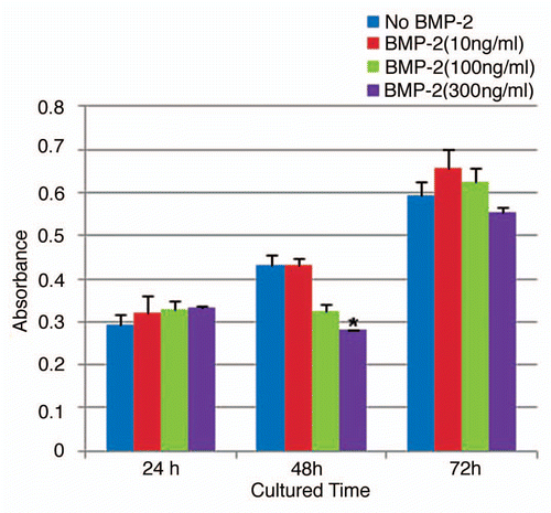
Figure 2 Effects of BMP-2 on ALDHbr cell growth. Freshly sorted ALDHbr cells were cultured for expansion and then inoculated in 96-well plates at a density of 1 × 10⁴ cells/well for 24 h. Cells were then cultured in 0.5% serum-containing medium for another 24 h after washing in phosphate-buffered saline. Cells were treated with 10, 100 or 300 ng/mL BMP-2 diluted in 0.5% serum-containing medium or vehicle control for 24, 48 and 72 h. Growth of the ALDHbr cells was significantly inhibited by the addition of 300 ng/mL of BMP-2 in the presence of 0.5% FBS for 48 h (p < 0.05). each experiment was performed three times; representative examples are shown.

Figure 3 BMP-2 downregulates expression of embryonic stem cell markers in human osteosarcoma tumorigenic ALDHbr cells. Relative quantitative mRNA expression of Oct3/4a, Nanog and Sox-2 genes in ALDHbr cells freshly isolated from OS99-1 xenografts treated with 300 ng/mL of BMP-2 for 48 h. Gene expression levels were normalized to β-actin. Oct3/4, Nanog and Sox-2 in ALDHbr cells were all consistently lower than those in control cells (*p < 0.05; **p < 0.001). each experiment was performed three times; representative examples are shown.
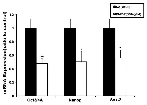
Figure 3 BMP-2 downregulates expression of embryonic stem cell markers in human osteosarcoma tumorigenic ALDHbr cells. Relative quantitative mRNA expression of Oct3/4a, Nanog and Sox-2 genes in ALDHbr cells freshly isolated from OS99-1 xenografts treated with 300 ng/mL of BMP-2 for 48 h. Gene expression levels were normalized to β-actin. Oct3/4, Nanog and Sox-2 in ALDHbr cells were all consistently lower than those in control cells (*p < 0.05; **p < 0.001). each experiment was performed three times; representative examples are shown.

Figure 4 BMP-2 upregulates expression of osteogenic markers in human osteosarcoma tumorigenic ALDHbr cells. Relative quantitative mRNA expression of Runx-2 and collagen type I genes in ALDHbr cells freshly isolated from OS99-1 xenografts treated with 300 ng/mL of BMP-2 for 48 h. Gene expression levels were normalized to β-actin. Runx-2 and collagen type I in ALDHbr cells were all consistently higher than those in control cells (*p < 0.05). Each experiment was performed three times; representative examples are shown.
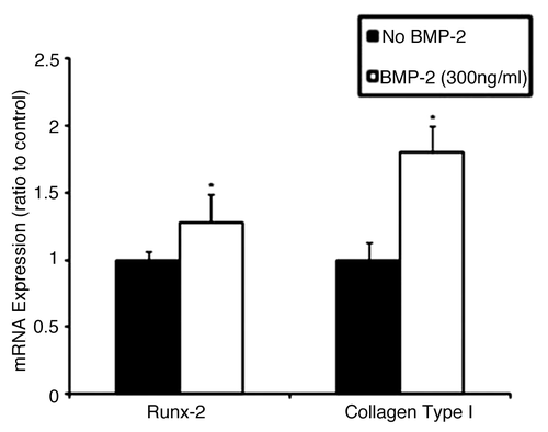
Figure 4 BMP-2 upregulates expression of osteogenic markers in human osteosarcoma tumorigenic ALDHbr cells. Relative quantitative mRNA expression of Runx-2 and collagen type I genes in ALDHbr cells freshly isolated from OS99-1 xenografts treated with 300 ng/mL of BMP-2 for 48 h. Gene expression levels were normalized to β-actin. Runx-2 and collagen type I in ALDHbr cells were all consistently higher than those in control cells (*p < 0.05). Each experiment was performed three times; representative examples are shown.

Figure 5 BMP-2 inhibits tumor formation by human osteosarcoma tumorigenic ALDHbr cells in vivo. (A and B) Representative tumor growth without BMP-2 treatment at the injection site in a NOD/SCID mouse. No significant tumor formation is seen at the injection site in BMP-2 treatment counterpart. (C) Representative hematoxylin and eosin staining of tumor generated from ALDHbr cells without BMP-2 treatment reveals presence of malignant cells showing characteristic osteosarcoma features, including nuclear atypia, extensive neovascularization and high mitotic activity (arrows). Large vesicle-like structures were Affi-Gel blue beads. (D) Injection site of ALDHbr cells treated with BMP-2 contained only residual large vesicle-like Affi-Gel blue beads, foreign-body giant cells (arrows) and fibrocytes.
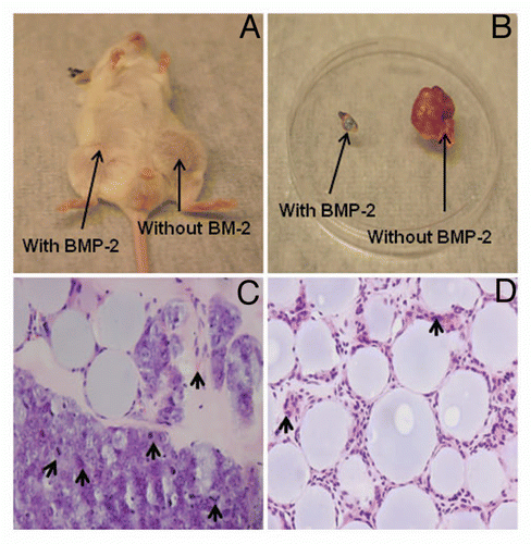
Figure 5 BMP-2 inhibits tumor formation by human osteosarcoma tumorigenic ALDHbr cells in vivo. (A and B) Representative tumor growth without BMP-2 treatment at the injection site in a NOD/SCID mouse. No significant tumor formation is seen at the injection site in BMP-2 treatment counterpart. (C) Representative hematoxylin and eosin staining of tumor generated from ALDHbr cells without BMP-2 treatment reveals presence of malignant cells showing characteristic osteosarcoma features, including nuclear atypia, extensive neovascularization and high mitotic activity (arrows). Large vesicle-like structures were Affi-Gel blue beads. (D) Injection site of ALDHbr cells treated with BMP-2 contained only residual large vesicle-like Affi-Gel blue beads, foreign-body giant cells (arrows) and fibrocytes.

Figure 6 Nuclear Ki-67 staining by immunohistochemistry. (A) Few positive cells were seen in the sections containing ALDHbr cells treated with 30 µg of BMP-2. Large vesicle-like structures were Affi-Gel blue beads. (B) Representative tissue sections of tumor formed by ALDHbr cells without BMP-2 treatment showed most cells were Ki-67 positive.
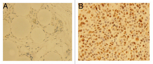
Figure 6 Nuclear Ki-67 staining by immunohistochemistry. (A) Few positive cells were seen in the sections containing ALDHbr cells treated with 30 µg of BMP-2. Large vesicle-like structures were Affi-Gel blue beads. (B) Representative tissue sections of tumor formed by ALDHbr cells without BMP-2 treatment showed most cells were Ki-67 positive.

Reprints and Corporate Permissions

Please note: Selecting permissions does not provide access to the full text of the article, please see our help page How do I view content?

To request a reprint or corporate permissions for this article, please click on the relevant link below:

Academic Permissions

Please note: Selecting permissions does not provide access to the full text of the article, please see our help page How do I view content?

Obtain permissions instantly via Rightslink by clicking on the button below:

If you are unable to obtain permissions via Rightslink, please complete and submit this Permissions form. For more information, please visit our Permissions help page.