Figures & data
Figure 1 Tramp prostate tumors and metastasis. (A) Large prostate ventral lobe tumors; normal seminal vesicles. (B) Metastatic lesions in lymph nodes (LN).
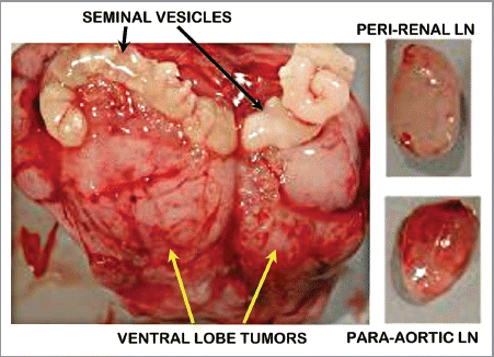
Figure 2 In vivo 3T 1H MRSI spectra taken from a region of normal peripheral zone and spectra taken from a region of prostate cancer and corresponding H&E stained sections identified on the corresponding T2 weighted MR image (center). 1H MRSI provides arrays of contiguous 1H MR spectra from throughout the prostate (white grid overlaid on MR image) and the spectra were selected to be localized within regions of healthy and cancerous tissues based on concordance of step-section histology of the resected prostate and T2 weighted MRI. Citrate is dramatically reduced in the Gleason 4+4 cancer as compared with healthy peripheral zone.
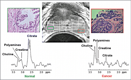
Figure 3 (A) Ex vivo 11.7T 1H HR-MAS spectra tissues taken from the normal murine prostatic ventral lobe, early stage TRAMP tumor, late stage TRAMP tumor, healthy human peripheral zone tissue and Gleason 4+3 prostate cancer and a metastatic lymph node in a TRAMP mouse. Note the appearance of citrate (4 peaks) is different than in the in vivo 1H MRSI spectra (two overlapping peaks). This is due to the increased chemical shift dispersion that occurs at 11.7T vs. 3T. (B) Quantification (Citrate/creatine peak area ratio) of the reduction in citrate on progression from normal to late stage and metastatic prostate cancer.
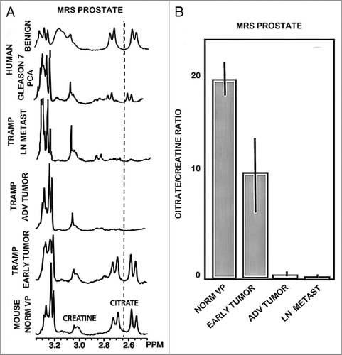
Figure 4 Dithizone zinc stain of prostate tissue sections. (A) Normal mouse prostate showing Zn-DTZ dark pigment formation due to presence of zinc. TRAMP tumors exhibit loss of zinc staining. (B) Our DTZ stain showing high Zn in normal peripheral zone glandular epithelium vs loss of Zn in peripheral zone adenocarcinoma. (C) DTZ staining of human prostate section which shows high Zn-DTZ in benign glandular epithelium and loss of zinc in malignancy (Gyorkey et al.Citation9).
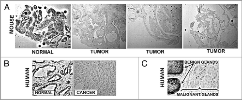
Figure 5 (A) ZIP1 transporter in normal mouse prostate and TRAMP tumors. ZIP1 IHC reveals the presence of the transporter in normal prostate acinus. The transporter is localized at the glandular epithelium basal membrane (blue arrows), and at the apical membrane (red arrows). There is no detectable transporter in the TRAMP tumors. (B) ZIP1 transporter in human prostate (taken from Franklin et al.Citation10). Shows same ZIP1 relationship as in (A).
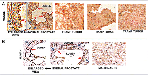
Figure 6 ZIP1 transporter western blot analysis of TRAMP C2 cells. 48 and 66 kDa bands are membrane-complexed forms of the transporter protein. 35 kDa is likely the free cytosolic protein.
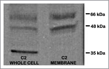
Figure 7 Effects of zinc treatment on TRAMP C2 cells. (A) Effect on cellular accumulation of zinc. (B) Effect on cell proliferation. (C) Effect on apoptosis.
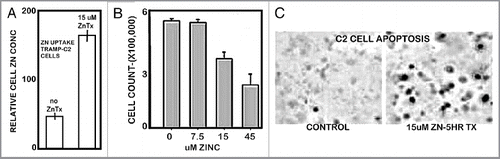
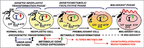
Figure 8 The ZIP1/zinc/citrate genetic/metabolic transformation concept of prostate carcinogenesis.