Figures & data
Figure 1. Screening of targeted human cancer cell line and tests of clinical samples. (A) PERP expression was assayed by immunobloting in some human cancer cell lines. SMMC7721, hepatocellular carcinoma; EC9706, esophageal carcinoma; Anip973, lung adenocarcinoma; 686/LN-1, oral squamous cell carcinoma lymph node metastasis; 686/LN-1-PERP, 686/LN-1 transfected with pcDNA3.1-PERP plasmid. (B) Four-paired samples of human lung adenocarcinoma were detected by PERP immunobloting. C, cancerous tissue of lung adenocarcinoma; N, normal lung tissue of the same patient.
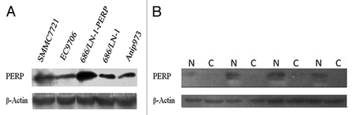
Figure 2. PERP overexpression inhibit lung cancer development in BALB/c nude mice. (A) PERP expression was detected in xenografts from N.S, Vector and PERP group, respectively. (B and C) Tumor growth curve (B) and weight (C) for Anip973 xenografts in mice undergoing PERP intervention. *p < 0.05, **p < 0.01. (D) H&E stainning of xenograft sections from N.S, Vector and PERP group, respectively. Arrows denote adenoid structure. Scale bar represents 100 μm.
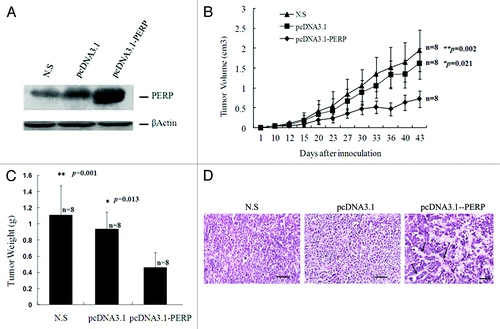
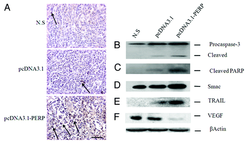
Figure 3. PERP induces apoptosis and downregulate VEGF. (A) TUNEL staining of Anip973 xenografts from N.S, Vector and PERP group, respectively. Arrows denote TUNEL positive tumor nuclei. Scale bar represents 100 μm. (B–F) Expression of Caspase 3, PARP, Smac, TRAIL and VEGF by immunoblotting were statistically significant in Anip973 xenografts from N.S, Vector and PERP group, respectively.