Figures & data
Figure 1 Sarcosphere size and efficiency reflect tumorigenic potential in osteosarcoma. (A) MNNG/HOS and 143B OS cell lines generates larger sarcospheres than Saos-2 and U-2 OS (*p < 0.01, size in microns, at least 100 spheres per group). (B) Representative sarcosphere derived from MNNG/HOS and U-2 OS. Bar 100 µm. (C) MNNG/HOS and 143B form more sarcospheres than Saos-2 and U-2 OS (SFE *p < 0.01, six samples/group in duplicate experiments). (D) Total number of spheres per well of MNNG/HOS and U-2 OS. Bar 500 µm. (E) 143B (top) has increased cell migration capacity than Saos-2 (bottom). The images at the beginning and the end of the 24 h incubation period were captured. The estimated migration of 143B cells was 2.7 times faster than Saos-2 cells (cell migration velocity (µm/h) 14.5 ± 0.85 vs. 5.3 ± 0.89, mean ± SD, p < 0.001, gap distance 600 µm, performed four times). (F) Metastatic osteosarcoma sample (OS1095) generates sarcospheres more efficiently than other primary bone tumor samples (SEF *p < 0.01, six samples/group in duplicate).
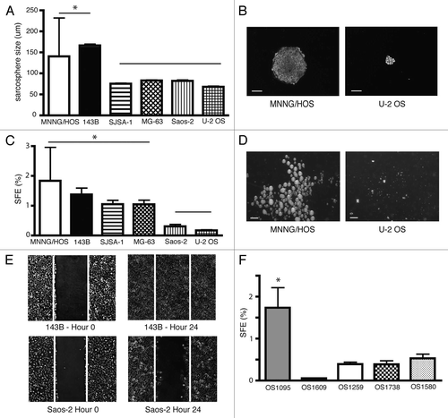
Figure 2 PKH26Hi ostesarcoma cells show increased self-renewal and divide less frequently. (A) PKH26Hi cells (black columns) generate twice as many sarcospheres than pKH26Lo cells (white columns) in three OS cell lines (SFE p < 0.01, at least three samples/group in duplicate experiments). (B) Only a minority of cells retain the PKH26 dye after serial sarcosphere passages (143B top row, primary sacosphere left, tertiary sarcosphere right; MNNG/HOS bottom row, primary sacosphere left, tertiary sarcosphere right). Bar 100 µm. (C) Representative FACS analysis shows PKH profile of cultured MNNG/HOS cell line (10,000 cells, PKH26Hi red, PKH26Lo gray, results were reproducible in at least two independent experiments). (D) Representative FACS analysis shows heterogeneous distribution of PKH26 dye on MNNG/HOS orthotopic bone tumors. Quiescence cells dilute less the membrane dye and appear brighter (10,000 cells, PKH26Hi red, PKH26Lo gray, results were reproducible in at least two independent experiments).
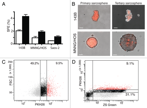
Figure 3 PKH26Hi osteosarcoma cells are more tumorigenic and generate bone tumors and lung metastasis. (A) Bone tumor (bright, left; ZsGreen, right) obtained after orthotopic injection of 1000 PKH26Hi cells. Bar 1 mm. (B) Pulmonary metastasis (bright, left; ZsGreen, right) are observed in some bone tumor bearing mice. Bar 1 mm. (C) sarcospheres obtained from bone tumors demonstrate the presence of PKH26 dye (bright, left; ZsGreen, right; PKH26, bottom) Bar 50 µm. (D) sarcospheres generated from pulmonary metastasis appeared to have an increased proportion of PKH26 cells (bright, left; ZsGreen, right; PKH26, bottom) Bar 50 µm.
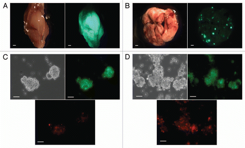
Figure 4 PKH26Hi cells have a gene signature distinct to the bulk of osteosarcoma cells in orthotopic bone tumors. Sixteen osteosarcoma samples obtained directly from orthotopic bone tumors were present on the Affymetrix U133A microarray platform. The heatmap represents the gene expression of PKH26Hi and PKH26Lo groups. The gene expression data are median centered with yellow being upregulated (82 genes) and blue being downregulated (44 genes).
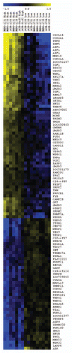
Table 1 Tumor-initiating cells were enriched in the PKH26Hi subpopulation