Figures & data
Figure 1 Structures of CP, Me-TPP+ and Mito-CP11.
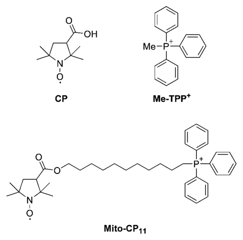
Figure 2 Antiproliferative effects of Mito-CP11 and CP in MCF-7 and MCF-10A cells. (A) MCF-7 and MCF-10A cells were treated with Mito-CP11 and CP (0.5 and 1 µM) for 48 h, and colonies formed were counted after 5 d (for MCF-7 cells) and 7 d (for MCF-10A cells). (B) The cell survival fractions calculated as described in the Materials and Methods section in Mito-CP11- and CP-treated MCF-7 and MCF-10A cells are shown. The plating efficiency calculated for MCF-7 and MCF-10A cells was 65 ± 13 and 38 ± 5, respectively. The error bars represent standard deviation and * symbol indicates a p value of less than 0.02 as determined by Student's t-test (n = 5).
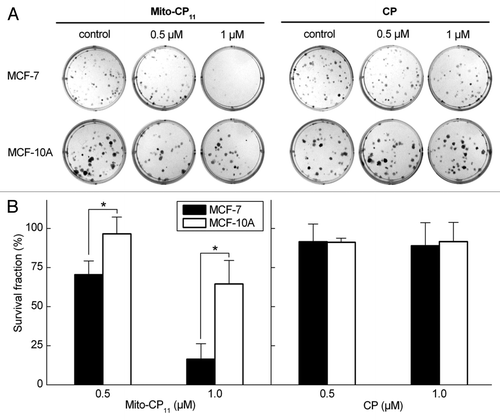
Figure 3 Antiproliferative effects of fluvastatin and Mito-CP11 in MCF-7 cells: Effect of mevalonate. (A) MCF-7 cells were treated with Mito-CP11 (0.5 µM) and fluvastatin (1 or 2.5 µM) in the presence and absence of mevalonate (20 µM) for 48 h, and the colonies counted after an additional 5 d. (B) Cell survival was measured under the same conditions as in (A). The survival fraction was calculated as described in the Materials and Methods section. The error bars represent standard deviation and * symbol indicates p value of less than 0.005 as determined by the Student's t-test (n = 9).
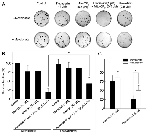
Figure 4 The effect of fluvastatin and Mito-CP11 on the extent of cell death in MCF-7 and MCF-10A cells. (A) MCF-7 and MCF-10A cells were treated with Mito-CP11 (0.5 µM) with or without fluvastatin (1 µM) and mevalonate (20 µM) for 48 h, and dead cells were monitored by staining with SYTOX Green. To measure the total cell number, cells were treated with digitonin (120 µM) when staining with SYTOX Green. Fluorescence intensity from cells grown in 96-well plate was measured using a plate reader (excitation wavelength, 485 nm and the emission wavelength 535 nm). (B) Fluorescence microscopy pictures were obtained using FITC filters under the same experimental cell culture conditions as in (A). Asterisks above a column indicate a statistical comparison between the indicated treatment and the control (*p < 0.05).
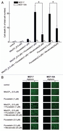
Figure 5 Uptake of Mito-CP11 into mitochondria. MCF-7 cells were treated with CP (5 µM) in the presence of Me-TPP+ and Mito-CP11 (5 µM) for 48 h. After treatment, cells were fractionated to isolate mitochondria and cytosol. CP and Mito-CP11 distributions in media, cytosol, mitochondria and cell lysate were determined using EPR. The EPR signals from different samples were normalized to the same concentration of protein.
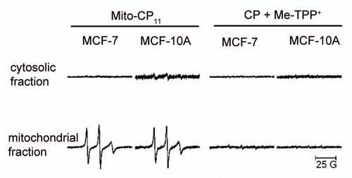
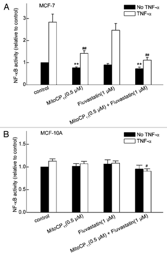
Figure 6 The effect of Mito-CP11 and fluvastatin on NFκB levels in MCF-7 and MCF-10A cells. The activity of NFκB in MCF-7 (A) and MCF-10A (B) cells was measured after incubating the cells for 48 h in the absence or presence of Mito-CP11 (0.5 µM) or fluvastatin (1 µM), followed by incubation in the absence or presence of TNFα (100 ng/ml, 3 h). NFκB activity was normalized to values in control cells that were incubated in the absence of Mito-CP11, fluvastatin and TNFα. Results are the mean ± SEM from three independent experiments conducted with triplicate samples. **p < 0.001 vs. control without TNFα; #p < 0.01, ##p < 0.001 vs. control with TNFα.