951
Views
18
CrossRef citations to date
0
Altmetric
Research Paper

Inhibition of metadherin sensitizes breast cancer cells to AZD6244

, , &
Pages 43-49 | Received 11 Jun 2011, Accepted 28 Nov 2011, Published online: 01 Jan 2012

Figures & data

Figure 1. Knockdown of MTDH sensitizes MDA-MB-231 cells to AZD6244. (A) Relative MTDH mRNA and protein levels in the breast cancer cell line MDA-MB-231 transfected with prpn and prpM vector, respectively. Error bars represent means ± SD of triplicate measurements. (B) Cell viability was evaluated using the MTT assay after treated cells with various concentration of AZD6244 for 96 h. Marked difference of survival fraction was observed between 231-prpn and 231-prpM cells (p = 0.005). Data shown are the means ± SD of three independent experiments. (C and D) Percentages of G0/G1 cells of each treated sample were measured using flow cytometric analysis for PI staining for cell cycle after 24 h treatment with AZD6244 0.5uM while the cells without treatment as control. (Data represent mean ± SD and results are representative of three independent experiments, *p < 0.01 vs. respect control). (E and F) 231-prpn and 231-prpM cells were treated with AZD6244 10 μM for 96 h and subjected to sub-G1 analysis using flow cytometric analysis with the cells without treatment as control. Cell death was measured by percentages of sub-G1 cells. Data represent mean ± SD and results are representative of three independent experiments, *p < 0.01 vs. respect control.
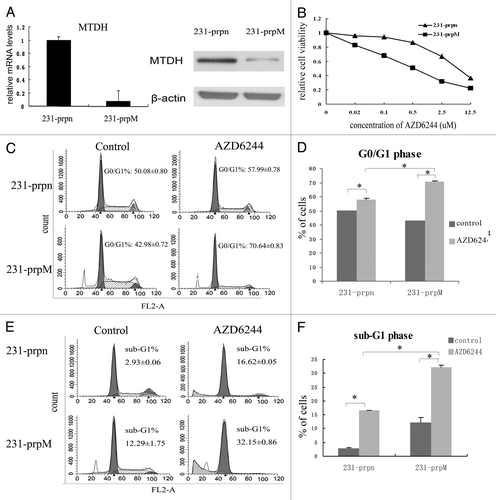
Figure 1. Knockdown of MTDH sensitizes MDA-MB-231 cells to AZD6244. (A) Relative MTDH mRNA and protein levels in the breast cancer cell line MDA-MB-231 transfected with prpn and prpM vector, respectively. Error bars represent means ± SD of triplicate measurements. (B) Cell viability was evaluated using the MTT assay after treated cells with various concentration of AZD6244 for 96 h. Marked difference of survival fraction was observed between 231-prpn and 231-prpM cells (p = 0.005). Data shown are the means ± SD of three independent experiments. (C and D) Percentages of G0/G1 cells of each treated sample were measured using flow cytometric analysis for PI staining for cell cycle after 24 h treatment with AZD6244 0.5uM while the cells without treatment as control. (Data represent mean ± SD and results are representative of three independent experiments, *p < 0.01 vs. respect control). (E and F) 231-prpn and 231-prpM cells were treated with AZD6244 10 μM for 96 h and subjected to sub-G1 analysis using flow cytometric analysis with the cells without treatment as control. Cell death was measured by percentages of sub-G1 cells. Data represent mean ± SD and results are representative of three independent experiments, *p < 0.01 vs. respect control.

Figure 2. MTDH knockdown increases expression of FOXO3a and promote its nuclear translocation. (A) The FOXO3a expression in protein level was examined by western blot, both ERK1/2 and AKT activity were measured by the level of p-ERK1/2 and p-AKT. (B) The expression of p-FOXO3a (Ser 294) was measured by western blot. The phosphorylation level of FOXO3a was measured by the ratio of p-FOXO3a/FOXO3a. (Data represent mean ± SD and results are representative of three independent experiments, *p < 0.01 vs. 231-prpn.) (C) The expression of FOXO3a in cytoplasma and nucleus was measured by western blot. (D and E) The efficiency of FOXO3a knockdown by siRNA in 231-prpn and 231-prpM cells was tested by western blot after transfected cells for 24 h. The changes of 231-prpM and 231-prpn cells sensitivity to AZD6244 was measured by MTT after treated cells for 96 h.
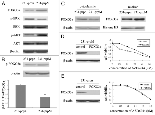
Figure 2. MTDH knockdown increases expression of FOXO3a and promote its nuclear translocation. (A) The FOXO3a expression in protein level was examined by western blot, both ERK1/2 and AKT activity were measured by the level of p-ERK1/2 and p-AKT. (B) The expression of p-FOXO3a (Ser 294) was measured by western blot. The phosphorylation level of FOXO3a was measured by the ratio of p-FOXO3a/FOXO3a. (Data represent mean ± SD and results are representative of three independent experiments, *p < 0.01 vs. 231-prpn.) (C) The expression of FOXO3a in cytoplasma and nucleus was measured by western blot. (D and E) The efficiency of FOXO3a knockdown by siRNA in 231-prpn and 231-prpM cells was tested by western blot after transfected cells for 24 h. The changes of 231-prpM and 231-prpn cells sensitivity to AZD6244 was measured by MTT after treated cells for 96 h.

Reprints and Corporate Permissions

Please note: Selecting permissions does not provide access to the full text of the article, please see our help page How do I view content?

To request a reprint or corporate permissions for this article, please click on the relevant link below:

Academic Permissions

Please note: Selecting permissions does not provide access to the full text of the article, please see our help page How do I view content?

Obtain permissions instantly via Rightslink by clicking on the button below:

If you are unable to obtain permissions via Rightslink, please complete and submit this Permissions form. For more information, please visit our Permissions help page.