1,354
Views
16
CrossRef citations to date
0
Altmetric
Research Paper

Combination of oncolytic adenovirus and dacarbazine enhances antitumor ability against uveal melanoma cells via cell cycle block

, , , , , & show all
Pages 77-84 | Received 05 Jul 2011, Accepted 16 Oct 2011, Published online: 15 Jan 2012

Figures & data

Figure 1. Sensitivity of human UM cell lines to adenovirus. (A) Infectivity of adenovirus Ad-GFP for human UM cell lines. Cells were infected with Ad-CMV-GFP adenovirus at an MOI of 20 pfu/cell, and GFP values were analyzed by flow cytometry. (B) Cytotoxicity of oncolytic adenovirus H101. Different cells were treated with H101 at an MOI of 50 pfu/cell and cell toxicity was determined by MTT assay 72 h post-infection. The data represent three independent experiments and the p values were determined by comparing VUP and SP6.5 vs. OM431 and OCM1. *p < 0.05, **p < 0.01
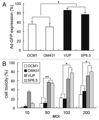
Figure 1. Sensitivity of human UM cell lines to adenovirus. (A) Infectivity of adenovirus Ad-GFP for human UM cell lines. Cells were infected with Ad-CMV-GFP adenovirus at an MOI of 20 pfu/cell, and GFP values were analyzed by flow cytometry. (B) Cytotoxicity of oncolytic adenovirus H101. Different cells were treated with H101 at an MOI of 50 pfu/cell and cell toxicity was determined by MTT assay 72 h post-infection. The data represent three independent experiments and the p values were determined by comparing VUP and SP6.5 vs. OM431 and OCM1. *p < 0.05, **p < 0.01

Figure 2. Cytotoxicity of combination treatment of H101 and DTIC on UM cells and normal cells. (A) morphological changes under light microscopy. SP6.5 cells were treated with DTIC, H101, and DTIC + H101, respectively. Original magnification × 200. (B) Cytotoxicity of DTIC, H101, and DTIC + H101 on SP6.5 (50 MOI + 5 µg/ml) and VUP cells (50 MOI + 2 µg/ml). Cells were subjected to the MTT assay at 24, 48, 72 and 96 h after treatment. (C) Human normal pigment epithelial cells ARPE-19 were treated exactly as above to evaluate the safety of the co-treatment. The results are representative of three independent experiments. *p < 0.05 compared with the DTIC or H101 treatment group.
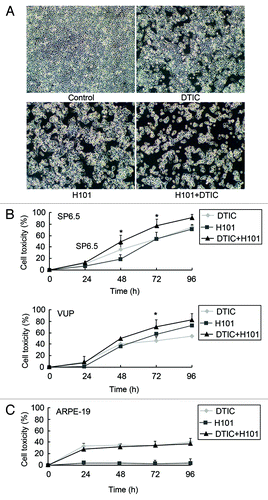
Figure 2. Cytotoxicity of combination treatment of H101 and DTIC on UM cells and normal cells. (A) morphological changes under light microscopy. SP6.5 cells were treated with DTIC, H101, and DTIC + H101, respectively. Original magnification × 200. (B) Cytotoxicity of DTIC, H101, and DTIC + H101 on SP6.5 (50 MOI + 5 µg/ml) and VUP cells (50 MOI + 2 µg/ml). Cells were subjected to the MTT assay at 24, 48, 72 and 96 h after treatment. (C) Human normal pigment epithelial cells ARPE-19 were treated exactly as above to evaluate the safety of the co-treatment. The results are representative of three independent experiments. *p < 0.05 compared with the DTIC or H101 treatment group.

Figure 3. Detection of apoptosis in SP6.5 cells. Apoptosis was measured by flow cytometry analysis 24 h after treatment with DTIC, H101, and DTIC + H101. The percentage numbers belongs to the two quadrants to the right. Results are representative of three independent experiments.
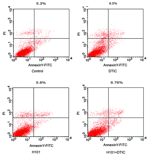
Figure 3. Detection of apoptosis in SP6.5 cells. Apoptosis was measured by flow cytometry analysis 24 h after treatment with DTIC, H101, and DTIC + H101. The percentage numbers belongs to the two quadrants to the right. Results are representative of three independent experiments.

Figure 4. Cell cycle distribution measurements and morphological changes of UM cells. (A) Cell cycle distribution of SP6.5 and VUP cells. Cell cycle analysis was performed at 48 and 72 h post-treatment with DTIC, H101, and DTIC + H101 by flow cytometry. ***p < 0.001 compared with G1 of control group, **p < 0.01 compared with G2 + S of control group, *p < 0.05 compared with G2 + S of control group in SP6.5 cells, *p < 0.05 compared with G1 of control group in VUP cells. The results are representative of three independent experiments. (B) Morphological changes of the nuclei SP6.5 cells were stained by DAPI 48 h after treatment with DTIC, H101 and DTIC+H101.
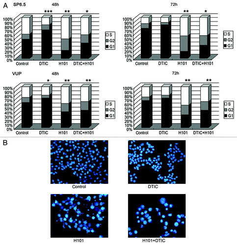
Figure 4. Cell cycle distribution measurements and morphological changes of UM cells. (A) Cell cycle distribution of SP6.5 and VUP cells. Cell cycle analysis was performed at 48 and 72 h post-treatment with DTIC, H101, and DTIC + H101 by flow cytometry. ***p < 0.001 compared with G1 of control group, **p < 0.01 compared with G2 + S of control group, *p < 0.05 compared with G2 + S of control group in SP6.5 cells, *p < 0.05 compared with G1 of control group in VUP cells. The results are representative of three independent experiments. (B) Morphological changes of the nuclei SP6.5 cells were stained by DAPI 48 h after treatment with DTIC, H101 and DTIC+H101.

Figure 5. Effect of co-treatment on adenoviral DNA replication in SP6.5 cells. (A) Viral early gene E1A expression was detected by q-PCR 24 h post-treatment with H101 or DTIC + H101. (B) Viral late gene Hexon expression was detected by q-PCR 72 h post-treatment with H101 or DTIC + H101. For comparison, the E1A and Hexon expression after H101 treatment was defined as 1. The results are representative of three independent experiments. (C) Western blot analysis of viral late protein Fiber expression 24, 48 and 72 h after DTIC, H101, and DTIC + H101 treatment.
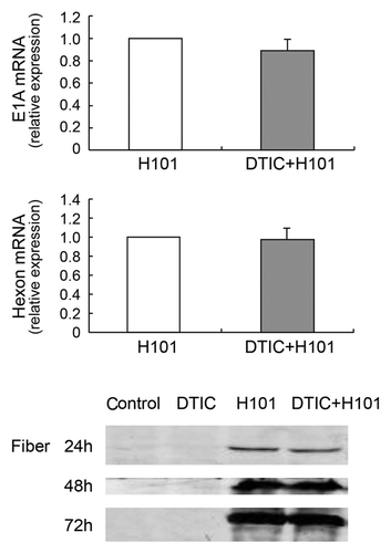
Figure 5. Effect of co-treatment on adenoviral DNA replication in SP6.5 cells. (A) Viral early gene E1A expression was detected by q-PCR 24 h post-treatment with H101 or DTIC + H101. (B) Viral late gene Hexon expression was detected by q-PCR 72 h post-treatment with H101 or DTIC + H101. For comparison, the E1A and Hexon expression after H101 treatment was defined as 1. The results are representative of three independent experiments. (C) Western blot analysis of viral late protein Fiber expression 24, 48 and 72 h after DTIC, H101, and DTIC + H101 treatment.

Reprints and Corporate Permissions

Please note: Selecting permissions does not provide access to the full text of the article, please see our help page How do I view content?

To request a reprint or corporate permissions for this article, please click on the relevant link below:

Academic Permissions

Please note: Selecting permissions does not provide access to the full text of the article, please see our help page How do I view content?

Obtain permissions instantly via Rightslink by clicking on the button below:

If you are unable to obtain permissions via Rightslink, please complete and submit this Permissions form. For more information, please visit our Permissions help page.