1,389
Views
11
CrossRef citations to date
0
Altmetric
Research Paper

Overexpression of centrosomal protein Nlp confers breast carcinoma resistance to paclitaxel

, , &
Pages 156-163 | Received 22 Sep 2011, Accepted 07 Nov 2011, Published online: 01 Feb 2012

Figures & data

Figure 1. Nlp locates in centrosome. (A) The identification of transfected cell lines: MCF-7 breast cancer cell line was transfected by pEGFP-C3 and pEGFP-Nlp respectively. Nlp clones and control were detected by protein gel blot analysis. β-Actin was used as an internal standard to indicate equal loading for each clone. (B) The localization of Nlp in centrosome was shown by immunofluoresent approach (red arrow marked bright green points).
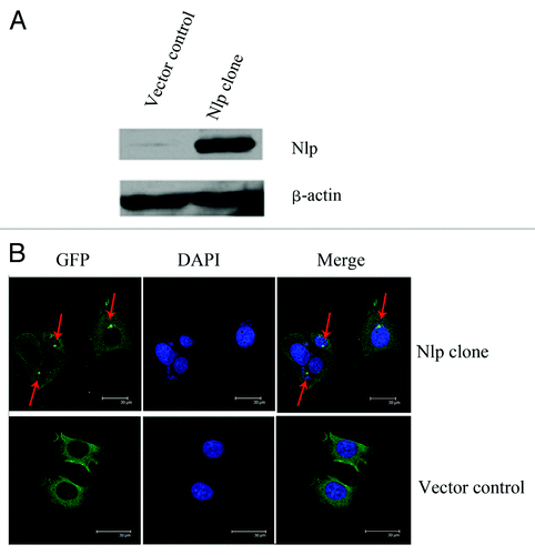
Figure 1. Nlp locates in centrosome. (A) The identification of transfected cell lines: MCF-7 breast cancer cell line was transfected by pEGFP-C3 and pEGFP-Nlp respectively. Nlp clones and control were detected by protein gel blot analysis. β-Actin was used as an internal standard to indicate equal loading for each clone. (B) The localization of Nlp in centrosome was shown by immunofluoresent approach (red arrow marked bright green points).

Figure 2. Nlp enhanced G2-M phase arresting but induced less Sub G1 phase in transfected cells. (A) Nlp overexpression facilitate resistance to paclitaxel in transfected breast cancer cells. Paclitaxel with different concentrations were administered in Nlp transfected cells and control and then analyzed by MTT approach. At least three independent experiments were done and the average values were calculated. (B) Nlp enhanced G2-M phase arresting in transfected breast cancer cells but induced less Sub G1 phase analyzed by FCM. (a-d) With different concentrations, transfected cells were treated by paclitaxel for 24 h, the percentages of G2-M phase cells and Sub G1 phase cells in two transfected cells were analyzed by FCM approach. (a) Quantitative analyses of G2-M phase cells, in which the assays were repeated at least three times. (b) A representative figure of cell cycle G2 analyses. (c) Quantitative analyses of Sub G1 phase cells, in which the assays were repeated at least three times. (d) A representative figure of Sub-G1 analyses.
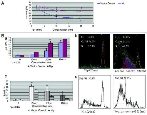
Figure 2. Nlp enhanced G2-M phase arresting but induced less Sub G1 phase in transfected cells. (A) Nlp overexpression facilitate resistance to paclitaxel in transfected breast cancer cells. Paclitaxel with different concentrations were administered in Nlp transfected cells and control and then analyzed by MTT approach. At least three independent experiments were done and the average values were calculated. (B) Nlp enhanced G2-M phase arresting in transfected breast cancer cells but induced less Sub G1 phase analyzed by FCM. (a-d) With different concentrations, transfected cells were treated by paclitaxel for 24 h, the percentages of G2-M phase cells and Sub G1 phase cells in two transfected cells were analyzed by FCM approach. (a) Quantitative analyses of G2-M phase cells, in which the assays were repeated at least three times. (b) A representative figure of cell cycle G2 analyses. (c) Quantitative analyses of Sub G1 phase cells, in which the assays were repeated at least three times. (d) A representative figure of Sub-G1 analyses.

Figure 3. Nlp protected breast cancer cells from cytotoxic apoptosis through suppressing activity of microtubule polymerization elicited by paclitaxel. Two transfected cells were treated by paclitaxel at different concentrations. Cells (attached cells plus those floating in the medium) were harvested and then were immunofluorescence stained by β-tubulin (red) antibody and DAPI (blue) to observe the changes of cytoskeleton after the paclitaxel treatment. The phenomena of microtubules polymerizing into bundles were clearer in the vector control compared with the Nlp transfected clone.
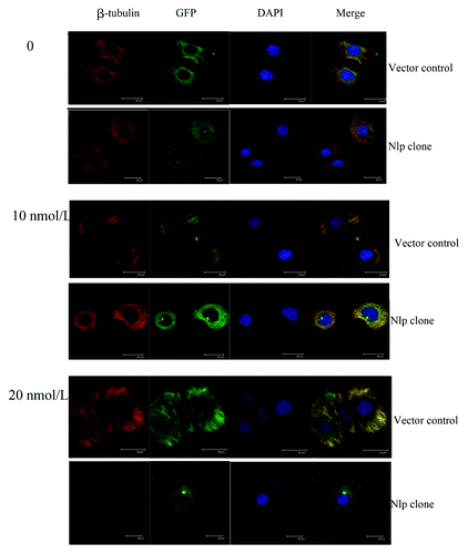
Figure 3. Nlp protected breast cancer cells from cytotoxic apoptosis through suppressing activity of microtubule polymerization elicited by paclitaxel. Two transfected cells were treated by paclitaxel at different concentrations. Cells (attached cells plus those floating in the medium) were harvested and then were immunofluorescence stained by β-tubulin (red) antibody and DAPI (blue) to observe the changes of cytoskeleton after the paclitaxel treatment. The phenomena of microtubules polymerizing into bundles were clearer in the vector control compared with the Nlp transfected clone.

Figure 4. Nlp might upregulate the expressions of Bcl-2 protein. Two transfected cells were treated by paclitaxel for different time followed by extracting their proteins. One hundred micrograms of whole-cell protein was used for protein gel blot analysis with anti-β-tubulin, anti-Bcl-2, anti-Plk1, anti-Cyclin B1 antibody as described in materials and methods. No differences of expressions of β-tubulin, Cyclin B1 and Plk1 in two transfected cells were shown, whereas, the expression of Bcl-2 increased significantly in Nlp transfected clone.
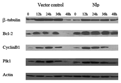
Figure 4. Nlp might upregulate the expressions of Bcl-2 protein. Two transfected cells were treated by paclitaxel for different time followed by extracting their proteins. One hundred micrograms of whole-cell protein was used for protein gel blot analysis with anti-β-tubulin, anti-Bcl-2, anti-Plk1, anti-Cyclin B1 antibody as described in materials and methods. No differences of expressions of β-tubulin, Cyclin B1 and Plk1 in two transfected cells were shown, whereas, the expression of Bcl-2 increased significantly in Nlp transfected clone.

Figure 5. Nlp expression in human breast cancer tissues. The samples derived from breast cancer patients were collected and subjected to immunohistochemical staining with anti-Nlp antibody, which indicates Nlp mainly expressed in cytoplasm.
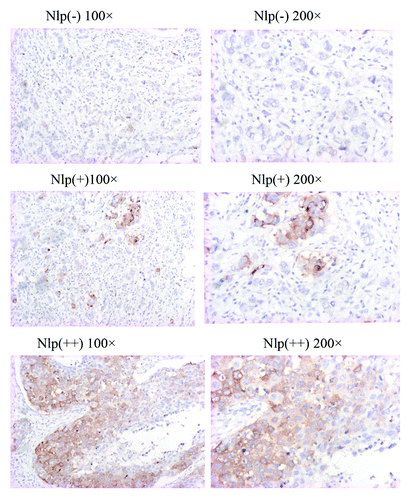
Figure 5. Nlp expression in human breast cancer tissues. The samples derived from breast cancer patients were collected and subjected to immunohistochemical staining with anti-Nlp antibody, which indicates Nlp mainly expressed in cytoplasm.

Table 1. Correlations between expression of Nlp protein and paclitaxel sensitivity

Table 2. Correlations between expression of Nlp protein and other prognostic factors in breast cancer patients

Reprints and Corporate Permissions

Please note: Selecting permissions does not provide access to the full text of the article, please see our help page How do I view content?

To request a reprint or corporate permissions for this article, please click on the relevant link below:

Academic Permissions

Please note: Selecting permissions does not provide access to the full text of the article, please see our help page How do I view content?

Obtain permissions instantly via Rightslink by clicking on the button below:

If you are unable to obtain permissions via Rightslink, please complete and submit this Permissions form. For more information, please visit our Permissions help page.