Figures & data
Figure 1 Effect of bortezomib and micelle-thiostrepton treatment on MDA-MB-231-luc tumor xenograft models. (A) Biodistribution of rhodamine-labeled micelle-thiostrepton complexes in tumor-bearing mice. High micelle-associated fluorescence is apparent in tumors and in the liver 4 h post-injection. Value for each organ represents an average of n = 2–4. (B) Anticancer effects of bortezomib (0.5 mg/kg) alone, micelle-thiostrepton treatment (30 mg/kg) alone, a combination of bortezomib and micelle-thiostrepton on MDA-MB-231 xenograft volumes, after five treatments over 15 d, n = 6–8. All averages were considered significant, and the t-test p value of the non-treated and the V+T-treated tumors was calculated to be 0.011. Error bars represent SEM. (C) Change in tumor-associated bioluminescence in animals bearing breast cancer xenografts after continuous treatment with a combination of complementary proteasome inhibitors over 15 d. Luciferase expression demonstrates that the sizes of tumors treated with bortezomib and thiostrepton were much smaller in size than the non-treated controls. (D) Change in animal weight during treatment schedule. Only the animals receiving the combination of proteasome inhibitors demonstrated slight weight loss, compared with non-treated and single drug-treated animals.
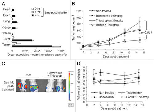
Figure 2 Combination treatment with bortezomib and micelle-thiostrepton induces cell death in MDA-MB-231-luc tumor xenografts, as determined by promotion of apoptotic markers. (A) Immunhistochemical analysis of four individual tumors (two from non-treated and two from combination treated groups) for cleaved caspase-3 expression (counterstained with hematoxylin). Cleaved-caspase-3 is evidently enhanced in combination-treated tumors, identified by the presence of larger numbers of dark, DAB-stained clusters. Control slides were incubated with rabbit IgG primary antibodies. Blue bars represent 100 µm (20 x). (B) Protein gel blotting of a panel of individual tumor homogenates (five of non-treated and five of combination-treated) against cleaved PARP and β-actin. Cleaved PARP expression is increased in tumors treated with a combination of thiostrepton and bortezomib.
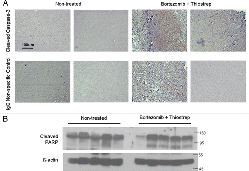
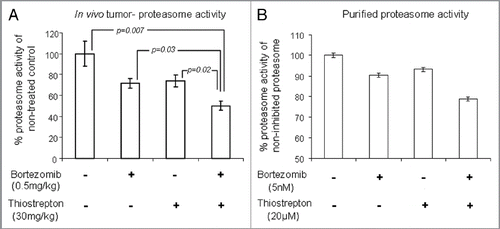
Figure 3 Combination of bortezomib and thiostrepton suppresses proteasome activity. (A) Proteasome activity assay showed that lysates from MDA-MB-231 tumors treated with a combination of proteasome inhibitors had decreased ability for proteasome substrate cleavage, compared with that from non-treated and single drug-treated tumors. Values represent averages of 6–8 tumors and error bars depict SEM p values are stated within the figure. (B) Proteasome activity assay showed that bortezomib and thiostrepton inhibit proteasome activities of purified proteasomes to a greater extent when used in combination.