Figures & data
Figure 1. Morphological characteristics of human triple-negative breast cancer (TNBC) and non-TNBC. (A) H&E staining of a TNBC and a non-TNBC. Tumor nests are composed of poorly differentiated small tumor cells. Necrosis in the center of a tumor nest is observed (black arrow). A number of tumor cells are undergoing mitosis (red arrows). IHC for ER, PR and hEGFR-2 indicate that the TNBC is negative for them, while the non-TNBC expresses PR and hEGFR-2. (B) IHC for p53, CK5/6 and CLU. CLU expression is localized in the cytoplasm of breast cancer cells, showing that the CLU identified in this case is sCLU. There is no expression of CLU in the non-TNBC. Ruler is 100 μm.
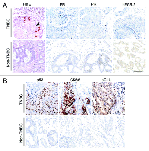
Table 2. Comparison of pathological and clinical features between triple-negative and non-triple-negative breast cancer cases
Figure 2. Survival of TN and non-TN breast cancer patients. (A) Overall survival rate of TN and non-TN breast cancer patients. At the end of the follow-up, 62.2% of TN patients survive, which is significantly less than that of non-TN patients. Kaplan-Meier survival analysis shows that the outcome of TN breast cancer is poorer than that of non-TN breast cancer (χ2 = 4.595, p = 0.032). (B) Effect of sCLU expression on TN breast cancer survival. In TN breast cancer patients, 70% of the sCLU-negative patients survive, whereas it is only 50% in the sCLU-positive group. (C) Effect of prechemotherapy on TN breast cancer survival. The survival rate of TN breast cancer patients pretreated with chemotherapy is nearly 90%, whereas it is less than 40% in the non-pretreatment group. Kaplan-Meier survival analysis shows that the difference is statistically significant (χ2 = 6.716, p = 0.010). (D) Effect of chemotherapy frequency after surgery on TN breast cancer survival. Approximately 80% of the patients that underwent less chemotherapy survive, whereas the survival rate of patients that underwent more chemotherapy is about 50%.
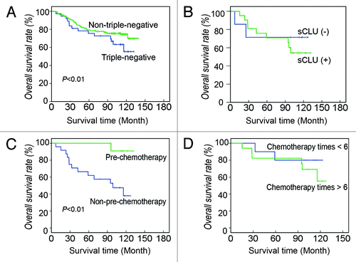
Table 3. Comparison of pathological and clinical features between sCLU-negative and positive triple-negative breast cancer cases
Figure 3. Morphological characteristics of TA2 spontaneous breast cancer. (A) TA2 spontaneous breast cancer is mostly composed of small round cells with few cytoplasms, and they form various tumor nests separated by well-developed stroma. The center of the solid tumor nest show necrosis (black arrow), and several cells are undergoing mitosis (red arrows). (B) TA2 spontaneous breast cancer metastasis in the lung. (C) TA2 spontaneous breast cancer metastasis in the liver. Tumors nest in the sinusoidal vessels of the liver; the black arrow indicates a necrotic area. (D) IHC for ER. Several tumor cells are positive for ER staining. (E) IHC for PR. No tumor cell expressing PR is found. (F) IHC for hEGFR-2. TA2 spontaneous breast cancer is negative for hEGFR-2. (G) IHC for p53. Mild to moderate p53 expression is detected in the tumor cells. (H) IHC for cyclin D1. Cyclin D1 expression is high in the neoplastic epithelial cells, indicating the high proliferative activity of breast cancer. (I) IHC for PCNA. Approximately 50% of the breast cancer cells express PCNA, indicating the high proliferation rate of TA2 breast cancer. Rulers are 100 μm.
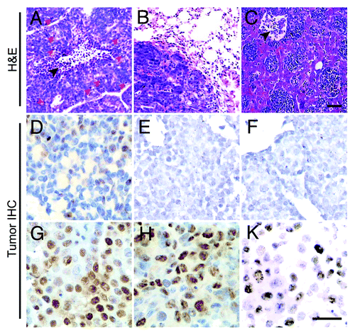
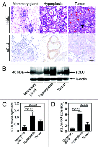
Figure 4. sCLU expression during TA2 spontaneous breast cancer progression. (A) H&E staining shows the morphology of a normal mammary gland, hyperplastic mammary glands, and breast cancer cells. Compared with the normal mammary gland, the number of cells and cell layers increase in the hyperplastic gland, and the cell and nucleus sizes also increase. IHC shows that the positive signal is localized in the cytoplasm, indicating that the protein is sCLU and not nCLU. A strong sCLU expression is detected in the mammary hyperplasia, whereas several mammary gland endothelial cells express sCLU. The sCLU expression in tumor cells is moderate. Ruler is 100 μm. (B) Western blot for clusterin (M-18). The expression of the 40 kDa sCLU is highest in the hyperplastic mammary epithelium, whereas the mammary epithelium of normal TA2 mice shows the lowest pre-sCLU expression. (C) Quantification of sCLU protein expression in the three groups (n = 4). The sCLU protein level in the normal mammary epithelium is significantly lower than that in the other groups. (D) sCLU mRNA expression among the three groups (n = 6). The hyperplastic mammary epithelium from TA2 mice with spontaneous breast cancer shows the highest sCLU mRNA expression among the three groups, whereas sCLU is the lowest in the mammary gland of normal TA2 mice. Spontaneous breast cancer expresses a middle level sCLU expression. Significant differences are found. The error bar means standard deviation (SD).