Figures & data
Figure 1. Protein expression levels of CaOV-3 and CHO-Ras stable transformants. CaOV-3 cells were stably transfected with an empty vector, 126/STOP, 223/STOP or 325/STOP, and expression levels of representative clones were shown by western blot. (A) Lane 1, empty vector, Lane 2, 126/STOP #12; Lane 3, 126/STOP #16; Lane 4, 223/STOP #8; Lane 5, 223/STOP #13; Lane 6, 325/STOP #21; Lane 7, 325/STOP #22. CHO-Ras cells were stably transfected with the same plasmids to get high protein expression levels. Conditioned media from representative CHO-Ras stable transformants were separated and stained with anti-IGF-IR α-subunit. (B) Lane 1, empty vector, Lane 2, 126/STOP #14; Lane 3, 126/STOP #16; Lane 4, 223/STOP #23; Lane 5, 223/STOP #26; Lane 6, 325/STOP #12; Lane 7, 325/STOP #15; Lane 8, 486/STOP #13.
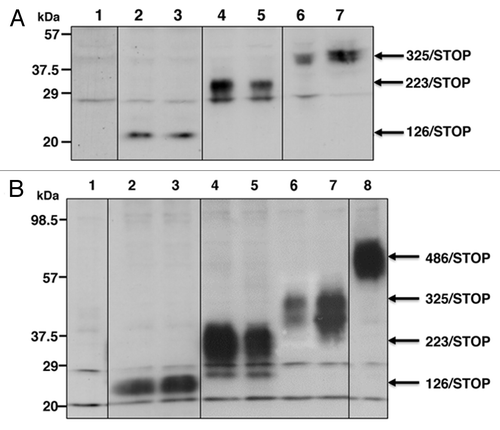
Figure 2. Cell growth in a monolayer culture. Representative CaOV-3 stable clones were plated at a density of 5 × 10⁴ cells in 6-well plates and incubated for 24 h in DMEM with 10% FBS. The medium was replaced either with SFM (closed columns), SFM+IGF-I (open columns) and SFM+10% FBS (striped columns). The number of cells was determined after an additional 48 h of incubation in each medium and is expressed as percentage increase. Columns, mean; bars, SD.
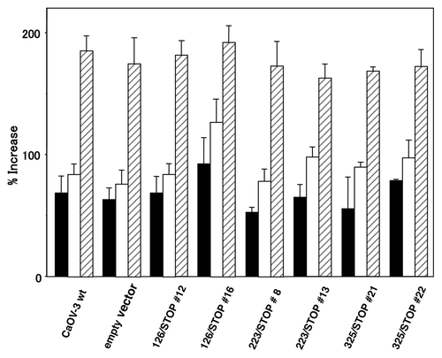
Figure 3. Colony formation in soft agar. Transfected CaOV-3 clones and CaOV-3 wild-type cells and an empty vector transfected clone were seeded at 3 × 10⁴ cells/35mm plate in 0.2% soft agar plates. Colonies grew > 200 μm in diameter were counted after 3 (closed columns), 4 (open columns) and 5 (striped columns) weeks incubation. *p < 0.01; **p < 0.001 vs. empty vector.
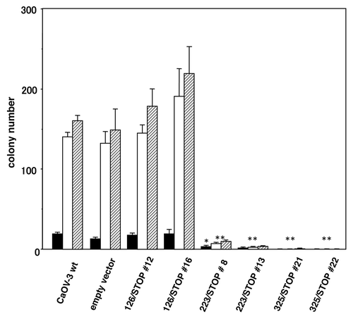
Figure 4. Tumor formation of transfected CaOV-3 clones in vivo. After 24 h of serum starvation, each clone (5 × 10⁶ cells) was resuspended in sterile PBS and injected s.c. in nude mice. Each point represents mean and SD from 5 mice. **p < 0.001 vs. empty vector.
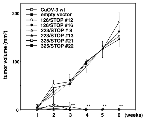
Figure 5. Evidence for apoptosis. After 24 h of serum starvation, various truncated IGF-IR transfected clones, wild-type CaOV-3 cells and an empty vector transfected clone were inoculated s.c. in nude mice. After 72 h inoculation, formed tumors were excised and stained with H&E or TUNEL.
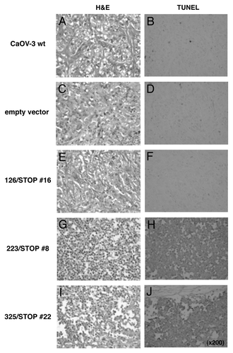
Figure 6. Bystander effects of the novel soluble IGF-I receptors to wild-type CaOV-3 cells. Conditioned media from CHO-Ras clones expressing various soluble IGF-IRs or the conditioned medium from an empty vector clone were prepared, then these conditioned media were mixed with the same amount of DMEM with 10% FBS for soft agar assay. Wild-type CaOV-3 cells at 3 × 10⁴ cells/35 mm plate were seeded in 0.2% agar (0.5% underlay) made of these mixed conditioned media. Definite colonies larger than 200 μm in diameter were counted after 1 week [open column] and 2 weeks [striped column]. *p < 0.01; **p < 0.001 vs. empty vector.
![Figure 6. Bystander effects of the novel soluble IGF-I receptors to wild-type CaOV-3 cells. Conditioned media from CHO-Ras clones expressing various soluble IGF-IRs or the conditioned medium from an empty vector clone were prepared, then these conditioned media were mixed with the same amount of DMEM with 10% FBS for soft agar assay. Wild-type CaOV-3 cells at 3 × 10⁴ cells/35 mm plate were seeded in 0.2% agar (0.5% underlay) made of these mixed conditioned media. Definite colonies larger than 200 μm in diameter were counted after 1 week [open column] and 2 weeks [striped column]. *p < 0.01; **p < 0.001 vs. empty vector.](/cms/asset/aa981e79-cb7f-484a-94f7-f2ff702c2a86/kcbt_a_10919609_f0006.gif)
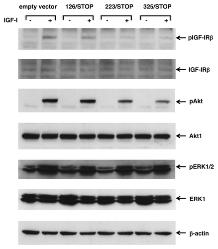
Figure 7. Transient expression of 233/STOP and 325/STOP in CaOV-3 cells downregulates IGF-I induced phosphorylation of IGF-IR and Akt. After transient transfection of empty vector or various soluble IGF-IRs, cells were kept in SFM for overnight, and then stimulated with 20 ng/ml IGF-I for 10 min at 37°C. The blots were probed with phosphorylation-specific antibodies, then stripped and re-probed with responsible antibodies. Expression levels of β-actin are shown as an internal control.
