Figures & data
Figure 1. TxnRd1 protein and activity levels in cells with different transformation status correlate with response to curcumin. (A) TxnRd1 protein levels were determined by immunoblot analysis in a panel of human head and neck cancer cell lines. FaDu, UPCISCC090, UPCISCC154 (both HPV+) SQ20B and JHU022. TxnRd1 level;s were also measured in the immortalized keratinocyte cell line HEK-001 and the leukoplakia derived line MSK = Leuk 1. β-Actin was used as a loading control. (B) Basal levels of whole-cell TxnRd activity was measured as nanomoles dihydrolipoate formed per milligram cell protein.
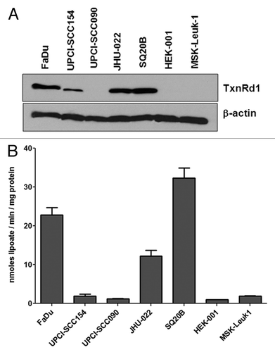
Figure 2. HPV- head and neck squamous carcinoma lines with TxnRd1 are sensitized to IR by curcumin. (A) Head and neck cell lines were treated with DMSO or 10 μM curcumin for 8 h followed by exposure to 0, 2, 4 or 6 Gy doses of IR. Survival was assessed by clonogenic assays. Points represent the average of a minimum of three independent experiments; bars ± SEM. Data were analyzed by pair wise comparison. * indicates a statistically significant difference in radiation sensitivity between curcumin treated and DMSO treated groups (p < 0.05). Values in boxes represent the dose modification factor observed at S.F. = 0.50. (B) Survival after a single dose of 2 Gy has been shown to predict intrinsic radiation sensitivity of tumor cells in vivo response. All HPV- HNSCC cell lines (FaDu, SQ20B and JHU-022, were significantly more resistant to 2 Gy of radiation then either HEK-001 or MSK-leuk-1 cells (*p < 0.05, t test). There was no difference in radiation sensitivity observed between the HPV+ cell lines and HEK-001 or MSK-Leuk 1. Pretreatment with curcumin, as indicated above, induced a significant increase in sensitivity in the three HPV- cell lines (* p < 0 0.05, t-test).
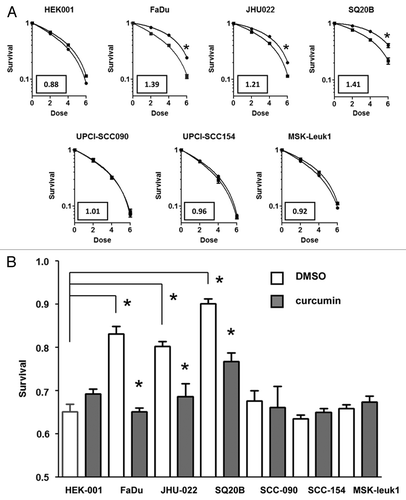
Figure 3. Growth of FaDu cells as an orthotopic intralingual model of HNSCC in nude mice and uptake of orally administered curcumin. (A) 10 μm cross-section of mouse tongue following H&E staining. (B) Immunohistochemical staining of a similar cross section for the proliferation marker Ki67. In both (A and B), the area labeled with T denotes tumor tissue; NS is normal stroma and NE is normal epithelium. (C) Uptake of curcumin into tumor tissues. Left panel shows a representative run for a tumor removed from an animal that was fed a 1% curcumin diet, (w/w) for 7 d, beginning 3 d after tumor implant. Right panel depicts a tumor-bearing mouse fed control lab chow. The peak that elutes at 40 min coincided with the peak obtained with the curcumin control. (D) Curcumin uptake was measured in three mice and the amount was determined by comparison to a curcumin standard. Error bars represent SEM values (p < 0.05; paired t-test). (E) TxnRd activity was measured in the tumor samples to insure that dietary curcumin was reaching the purported target in 3 mice fed 1% curcumin chow compared with 3 mice fed the control chow. Error bars represent SEM values (p < 0.05; paired t-test).
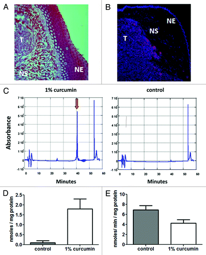
Figure 4. Effect of curcumin feeding with IR on orthotopic FaDu tumor growth was measured by bioluminescent image analysis. (A) FaDu cells transfected with CMV luciferase (FaDu-CMV-luci) were pretreated with 10 μM curcumin or DMSO for 8 h and then exposed to 0, 2, 4 or 6 Gy of radiation. Survival was measured by the clonogenic assay. The DMF = 1.42 for FaDu CMV-Luci cells, was similar to that of the parental FaDu cell line (). (B) Representative image from two groups of mice taken 10 d after completing 3 fractions of 2 Gy given at 48 h intervals. Left panel: from the group fed 1% curcumin chow; right panel: mice from the control chow fed group. (C) Tumor growth rates from each group of mice (10 mice/group at start of experiment) (control diet, 1% curcumin diet, 2Gy x 3 ionizing radiation alone or curcumin + ionizing radiation) based on twice-weekly bioluminescent imaging. Error bars represent SEM values from surviving mice at time of measurement. (D) Volume doubling time was calculated froom the time of the second pre-irradiation bioluminescent image intensity, obtained on day 7. Interaction was assessed by repeated measures ANOVA (p < 0.0001). The doubling time for the combined curcumin and radiation group was significantly larger than the unirradiated and irradiated mice on the control diet, and the curcumin-fed mice without radiation the radiation only treatment. Box represents minimum and maximum values for each group of animals with the median indicated by the interior bar. Error bars represent 95% confidence limits.
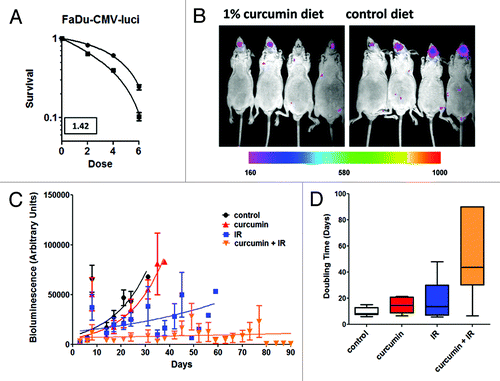
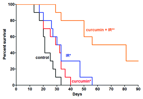
Figure 5. Effect of combined curcumin and IR on animal survival. Animals were removed from the study and euthanized when their body mass decreased by > 20% based on their weight at beginning of experiment. There was a statistical difference in mean survival between the curcumin alone or IR alone-treated groups when compared with the untreated animals (*p = 0.05). The difference in mean survival between the combined treatments group and all other groups was highly significant (**p = 0.0001). Notably, 3 animals from the combined treatment group survived beyond the last data point collected at day 90.