975
Views
3
CrossRef citations to date
0
Altmetric
Clinical Study

Increased frequency of circulating invariant natural killer T cells in malignant pleural mesothelioma patients

, , , , &
Pages 702-711 | Received 22 Dec 2011, Accepted 29 Apr 2012, Published online: 11 Jun 2012

Figures & data

Table 1. Characteristics of MPM patients and healthy volunteers

Figure 1. Flow cytometric analysis of iNKT cells and subsets from MPM patients and HV. PBMC were stained with anti-CD3 and anti-Vα24Vβ11 mAb. (A) Lymphocytes were gated on a side scatter (SSC)-forward scatter (FSC) dot plot. (B) In the lymphocyte gate, iNKT cells were identified as CD3+Vα24Vβ11+ double-positive cells. (C) Phenotypic analysis of iNKT cells was performed by CD4 and CD8 staining with anti-CD4 and anti-CD8 mAb. (D) A representative mean fluorescence intensity (MFI) histogram of CD3 expression on gate of lymphocytes. Representative flow cytometric dot plots of a HV (E) and a MPM patient (F); iNKT cell percentages are also shown in each panel. (G) The frequency of iNKT cells detected in the lymphocyte gate of MPM patients (n = 30; black bar) and HV (n = 22; unfilled bar). (H) Analysis of the frequency of iNKT cell subsets of MPM patients (black bars) and HV (unfilled bars). Results are expressed as mean ± SEM.
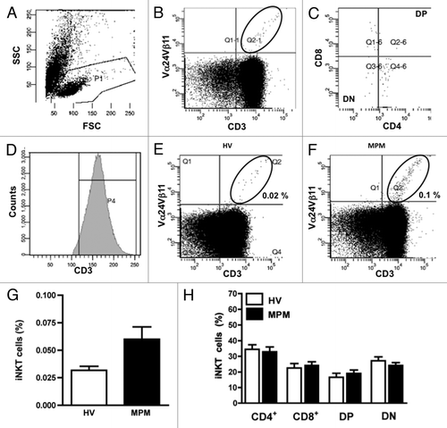
Figure 1. Flow cytometric analysis of iNKT cells and subsets from MPM patients and HV. PBMC were stained with anti-CD3 and anti-Vα24Vβ11 mAb. (A) Lymphocytes were gated on a side scatter (SSC)-forward scatter (FSC) dot plot. (B) In the lymphocyte gate, iNKT cells were identified as CD3+Vα24Vβ11+ double-positive cells. (C) Phenotypic analysis of iNKT cells was performed by CD4 and CD8 staining with anti-CD4 and anti-CD8 mAb. (D) A representative mean fluorescence intensity (MFI) histogram of CD3 expression on gate of lymphocytes. Representative flow cytometric dot plots of a HV (E) and a MPM patient (F); iNKT cell percentages are also shown in each panel. (G) The frequency of iNKT cells detected in the lymphocyte gate of MPM patients (n = 30; black bar) and HV (n = 22; unfilled bar). (H) Analysis of the frequency of iNKT cell subsets of MPM patients (black bars) and HV (unfilled bars). Results are expressed as mean ± SEM.

Figure 2. The frequency of circulating iNKT cells according to histological MPM subtypes. Scatter plot of iNKT frequencies in HV and biphasic- (B), epithelioid- (E) or sarcomatoid- (S) MPM patients. Horizontal bar is the median value for each group; *p < 0.05; **p < 0.01.
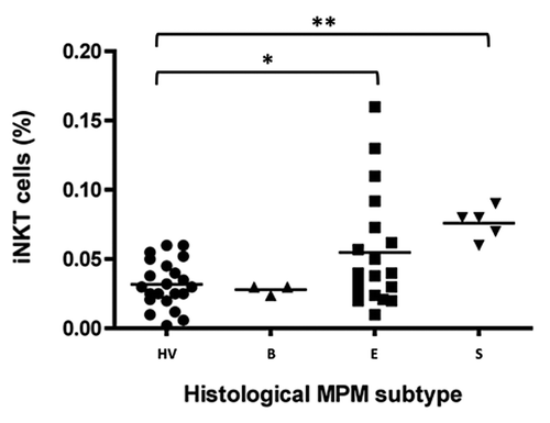
Figure 2. The frequency of circulating iNKT cells according to histological MPM subtypes. Scatter plot of iNKT frequencies in HV and biphasic- (B), epithelioid- (E) or sarcomatoid- (S) MPM patients. Horizontal bar is the median value for each group; *p < 0.05; **p < 0.01.

Figure 3. The frequency of iNKT cell subsets according to histological MPM subtypes. Analysis of the frequency of iNKT cell subsets was performed by CD4 and CD8 staining with anti-CD4 and anti-CD8 mAb. The values are expressed as percentage of iNKT cells expressing the molecules over the total iNKT cells. Data are the mean ± SEM; *p < 0.05.
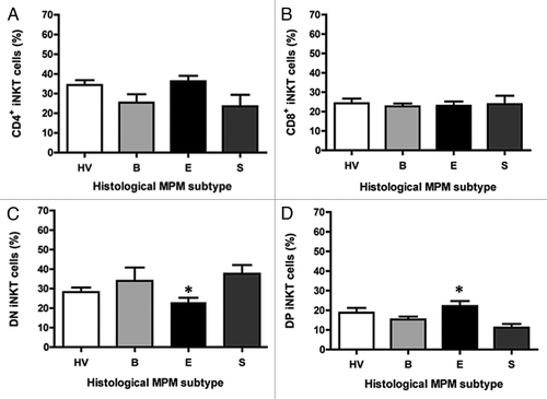
Figure 3. The frequency of iNKT cell subsets according to histological MPM subtypes. Analysis of the frequency of iNKT cell subsets was performed by CD4 and CD8 staining with anti-CD4 and anti-CD8 mAb. The values are expressed as percentage of iNKT cells expressing the molecules over the total iNKT cells. Data are the mean ± SEM; *p < 0.05.

Figure 4. The frequency of iNKT cells and iNKT cell subsets in epithelioid and sarcomatoid MPM patients belonging to a certain clinical stage. Scatter plot of the iNKT cell frequency in HV and patients with epithelioid (A) or sarcomatoid (B) MPM at different clinical stages. Horizontal bar is the median value for each group. **p ≤ 0.01. (C and D) Analysis of the frequencies of iNKT cell subsets was performed by CD4 and CD8 staining with anti-CD4 and anti-CD8 mAb. The values are expressed as percentage of iNKT cells expressing the molecules over the total iNKT cells. Data are the mean ± SEM.
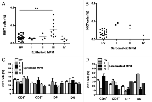
Figure 4. The frequency of iNKT cells and iNKT cell subsets in epithelioid and sarcomatoid MPM patients belonging to a certain clinical stage. Scatter plot of the iNKT cell frequency in HV and patients with epithelioid (A) or sarcomatoid (B) MPM at different clinical stages. Horizontal bar is the median value for each group. **p ≤ 0.01. (C and D) Analysis of the frequencies of iNKT cell subsets was performed by CD4 and CD8 staining with anti-CD4 and anti-CD8 mAb. The values are expressed as percentage of iNKT cells expressing the molecules over the total iNKT cells. Data are the mean ± SEM.

Figure 5. Intracellular cytokine expressions in iNKT cells. After stimulation of PBMC from HV and MPM patients with PMA/Io for 4 h, the cells were stained with anti-Vα24Vβ11 mAb followed by intracellular IFN-γ or IL-4 staining. (A) Representative dot plots displaying iNKT cell gates of unstimualted and PMA/Io-activated samples from a HV and a MPM patient. Percentages of IFN-γ and IL-4 positive cells in MPM patients (B and C) and in different MPM subtypes (D and E) compared with HV. Data are the mean ± SEM.
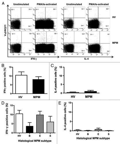
Figure 5. Intracellular cytokine expressions in iNKT cells. After stimulation of PBMC from HV and MPM patients with PMA/Io for 4 h, the cells were stained with anti-Vα24Vβ11 mAb followed by intracellular IFN-γ or IL-4 staining. (A) Representative dot plots displaying iNKT cell gates of unstimualted and PMA/Io-activated samples from a HV and a MPM patient. Percentages of IFN-γ and IL-4 positive cells in MPM patients (B and C) and in different MPM subtypes (D and E) compared with HV. Data are the mean ± SEM.

Reprints and Corporate Permissions

Please note: Selecting permissions does not provide access to the full text of the article, please see our help page How do I view content?

To request a reprint or corporate permissions for this article, please click on the relevant link below:

Academic Permissions

Please note: Selecting permissions does not provide access to the full text of the article, please see our help page How do I view content?

Obtain permissions instantly via Rightslink by clicking on the button below:

If you are unable to obtain permissions via Rightslink, please complete and submit this Permissions form. For more information, please visit our Permissions help page.