Figures & data
Figure 1. HO-3867 inhibits BRCA1 ovarian cancer cell proliferation. (A) Clonogenic assay of BRCA1-mutated ovarian cancer cells treated with increasing concentrations of HO-3867 for 24 h (1, 5, 10 and 20 µM). There is a significant decrease in the percentage of cell colonies in a dose response progression (left panel). (B) After treatment with increasing concentrations of HO-3867 (1, 5, 10, 20 µM) at serially sequential times (12, 24, 48 h) cells were collected and counted. Cell counts were significantly decreased starting with the 5 µM concentration. After 24 h treatment with 20 µM there were no viable cells remaining. (C) Conformation of decreased proliferation was performed with immunohistochemistry staining with DAPI. This demonstrated that cells treated with 10 µM of HO-3867 showed a significant decrease in the number of proliferating cells. (D) MTT assay of four ovarian cancer cell lines (A2780, A2780R, SKOV3 and BRCA1-mutated cells) treated with HO-3867 or STATTIC. While all cell lines showed significant decreased viability, this was most observed in the BRCA1-mutated cells. (E) Of interest HO-3867 showed more of an effect than the potent STAT3 inhibitor STATTIC. E, MTT assay of BRCA1-mutated cells with different treatments [Curcumin, cisplatin (CP), PARP-inhibitor (ABT), STATTIC and HO-3867]. While all treatments decreased cell viability the most dramatic effect was seen in the HO-3867 treatment group.
![Figure 1. HO-3867 inhibits BRCA1 ovarian cancer cell proliferation. (A) Clonogenic assay of BRCA1-mutated ovarian cancer cells treated with increasing concentrations of HO-3867 for 24 h (1, 5, 10 and 20 µM). There is a significant decrease in the percentage of cell colonies in a dose response progression (left panel). (B) After treatment with increasing concentrations of HO-3867 (1, 5, 10, 20 µM) at serially sequential times (12, 24, 48 h) cells were collected and counted. Cell counts were significantly decreased starting with the 5 µM concentration. After 24 h treatment with 20 µM there were no viable cells remaining. (C) Conformation of decreased proliferation was performed with immunohistochemistry staining with DAPI. This demonstrated that cells treated with 10 µM of HO-3867 showed a significant decrease in the number of proliferating cells. (D) MTT assay of four ovarian cancer cell lines (A2780, A2780R, SKOV3 and BRCA1-mutated cells) treated with HO-3867 or STATTIC. While all cell lines showed significant decreased viability, this was most observed in the BRCA1-mutated cells. (E) Of interest HO-3867 showed more of an effect than the potent STAT3 inhibitor STATTIC. E, MTT assay of BRCA1-mutated cells with different treatments [Curcumin, cisplatin (CP), PARP-inhibitor (ABT), STATTIC and HO-3867]. While all treatments decreased cell viability the most dramatic effect was seen in the HO-3867 treatment group.](/cms/asset/e9cda24b-3a42-4bf7-b8f8-7de34c7bdcd5/kcbt_a_10920559_f0001.gif)
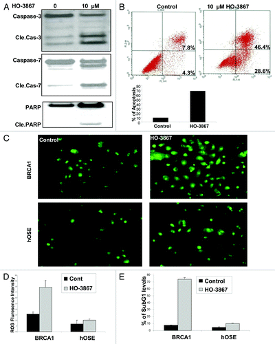
Figure 2. Effect of HO-3867 on apoptosis in BRCA1 ovarian cancer cells. (A) Representative western blot showing a significant increase in cleaved caspase -3, -7 and cleaved PARP after treatment with HO-3867. (B) Flow cytometry with propidium iodide and annexin V. Treatment with HO-3867 showed a significant increase in the number of cells in apoptosis from controls (75.0% vs 12.1%, respectively). Graphical representation of flow cytometry results shown. (C and D) Measurement of intracellular ROS in BRCA1-mutated cancer cells and normal hOSE cells. At baseline there is an increased measure of ROS in BRCA1 cancer cells vs. controls. After treatment with 10uM HO-3867 for 24 h, a significant increase in the number of cells with ROS was detected in BRCA1 cells. This observation, however, was not seen in hOSE cells showing HO-3867 was selective against the BRCA1-mutated cancer cell line while sparing normal, non-cancerous cells. (E) Graphical results from a subG1 analysis after treatment with HO-3867 for 24 h. After treatment, a significant increase in the number of BRCA1 cells remained in the subG1 phase.

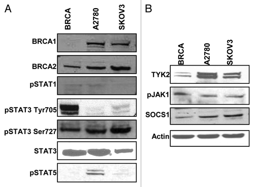
Figure 3. Activation of the STAT pathway in BRCA1-mutated ovarian cancer cells. (A) BRCA1, A2780 and SKOV3 cells were cultured and collected for western blot for expression of proteins in the STAT family. There was minimal expression of STAT1 or STAT5 (A2780 cells minimally expressed STAT5). All cell lines uniformily expressed STAT3 and the activated form pSTAT3 Ser727, however, only BRCA1 cells showed expression of pSTAT3 Tyr705. (B) Representative western blots for protein expression in the STAT3 pathway. BRCA1 cells showed less expression of TYK2 and SOCS1. Both pJAK1 and JAK2 were consistently expressed in all lines.

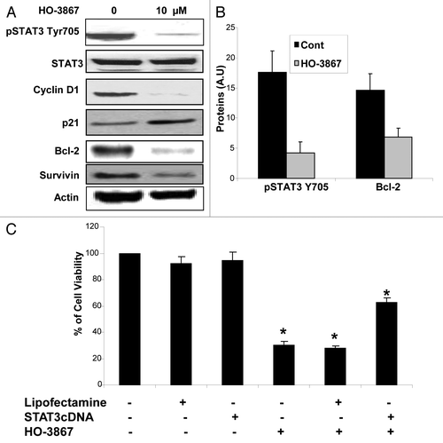
Figure 4. Effect of HO-3867 on STAT pathway protein expression. (A) BRCA1 cells were treated with 10uM of HO3867 for 24 h and protein expression analyzed via western blot. There was a significant decrease in the expression of activated pSTAT3 Tyr705, the downstream prosurvival proteins pAKT and Cyclin D1, as well as anti-apoptotic proteins Bcl-2 and Survivin. Increased expression of the cell cycle regulator p21 was seen as well. (B) Graphical representation confirms decrease of pSTAT3705 and Bcl-2 expression after treatment with 10uM HO-3867 for 24 h. (C) STAT3 overexpression leads to HO-3867 resistance. BRCA1-mutated cells were transfected with STAT3 cDNA to see if overexpression of STAT3 could lead to resistance of HO-3867 on cell viability. Cells that were treated with 10uM of HO-3867 with only the lipofectamine vector showed identical loss in cell viability as cells treated with HO-3867 alone. However, HO-3867-treated cells that were transfected with STAT3 cDNA had a significant increase in viability from those cells that were not transfected. Although there was improved survival there was still a significant decrease in viability from control cells showing the potency of HO-3867 as a STAT inhibitor.

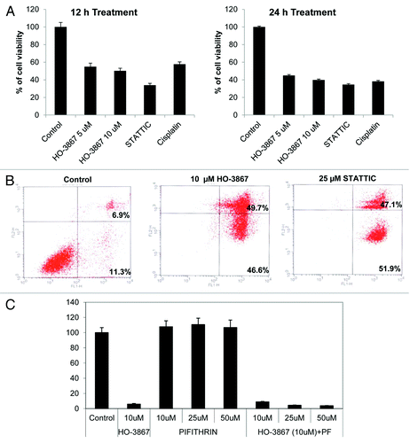
Figure 5. Effect of HO-3867 on viability and apoptosis in BRCA1-mutated breast cancer cells. (A) MTT assay of BRCA1-mutated breast cancer cell line (SUM149) treated with 5 or 10 μM HO-3867, 25 μM STATTIC or 10 μg/mL cisplatin for 12 or 24 h. Cells showed a significant decreased in viability as early as 12 h. By 24 h all treatments decreased viability by 50–60 percent. (B) Flow cytometry with propidium iodide and annexin V. Treatment with 10 μM HO-3867 and 25 μM STATTIC showed a significant increase in the number of cells in apoptosis from controls (96.3% and 99% vs 18.2%, respectively). Graphical representation of flow cytometry results shown. (C) Inhibition of p53 by pifithrin did not reverse the STAT3 inhibitor induced apoptosis. BRCA1 mutated ovarian cancer cells were pretreated with the pifithrin 10, 25 and 50 µM for 1 h and then treated with HO-3867 and STATTIC for 24 h. Cell viability was analyzed by MTT assay. STATTIC data not shown.
