Figures & data
Table 1. Anaplastic vs. papillary thyroid carcinoma: Immunohistochemical results
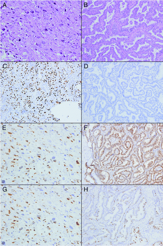
Figure 1. Hematoxylin and eosin stained sections of anaplastic (A) and papillary (B) thyroid carcinoma. ATC (C) showed a significant increase in staining for p53 as compared with PTC (D). Both ATC (E) and PTC (F) showed diffuse positive staining for cyclin D1. Although not statistically significant, ATC (G) was less likely to be p21 positive than PTC (H). Original magnification 200 x for all images.