Figures & data
Figure 1. A 54-y-old female patient with recurrent gliomas. (A) CT images before 125I seed implantation treatment showed that brain tumor of left posterior parietal region was 30 mm × 40 mm × 50 mm. (B) outlined the planning target volume (PTV) with computerized treatment planning system (TPS) before 125I seed implantation to determine the needle insertion and dose distributions. (C) needle insertion during 125I seed implantation operation. (D) seed distribution after 125I seed implantation treatment. E: verification of TPS immediately after 125I seed implantation treatment. (F) dose volume histogram of PTV of pre-operation TPS. (G) dose volume histogram of PTV of post-operation TPS verification.
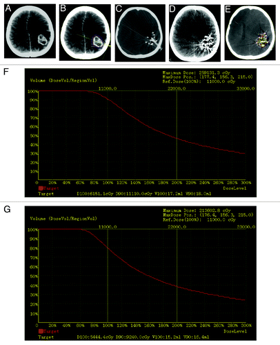
Figure 2. A 68-y-old male patient with recurrent gliomas. (A) CT images before 125I seed implantation treatment showed that brain tumor of left anterior parietal region was 25 mm × 30 mm × 40 mm. (B) needle insertion during 125I seed implantation operation. C:two months after 125I seed implantation treatment. (D) one year after 125I seed implantation treatment.
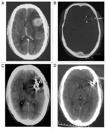
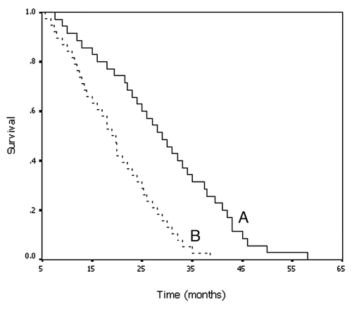
Figure 3. Kaplan-Meier curve shows overall survival rates for patients who received 125I implantation (A) and for those who underwent conventional chemoradiation therapy (B).