1,022
Views
47
CrossRef citations to date
0
Altmetric
Research Paper

Stromal galectin-1 expression is associated with long-term survival in resectable pancreatic ductal adenocarcinoma

, , , , , , , , , & show all
Pages 899-907 | Received 06 Feb 2012, Accepted 22 May 2012, Published online: 01 Aug 2012

Figures & data

Table 1. Differentially expressed proteins associated with long-term survivorship in pancreatic ductal adenocarcinoma

Figure 1. Identification of overexpression of galectin-1 in PDAC from STS in comparison to VLTS. The MS/MS spectrum leading to identification of a unique peptide (SFVLNLGK) from galectin-1 is displayed. The insert shows the iTRAQ reporting peaks of 114 and 117, representing STS and VLTS samples, respectively. The ratio of galectin-1 expression in STS relative to VLTS was calculated to be 1.91.
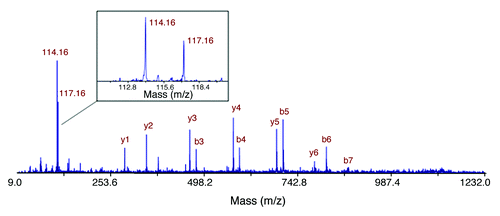
Figure 1. Identification of overexpression of galectin-1 in PDAC from STS in comparison to VLTS. The MS/MS spectrum leading to identification of a unique peptide (SFVLNLGK) from galectin-1 is displayed. The insert shows the iTRAQ reporting peaks of 114 and 117, representing STS and VLTS samples, respectively. The ratio of galectin-1 expression in STS relative to VLTS was calculated to be 1.91.

Figure 2. Galectin-1 staining in VLTS and STS in validation cohort 1 (Johns Hopkins cohort). Galectin-1 IHC of invasive pancreatic cancer displaying strong staining (3+, brown color) in desmoplatic stromal cells from a patient with short-term survival (A) and absent immunostaining in a VLTS patient (B). Distribution of galectin-1 staining from short-term survivors and very long-term survivors (C). ROC curve analysis displaying an ROC of 0.75, and 64% sensitivity and 90% specificity in predicting very long-term survival (D).
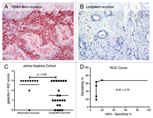
Figure 2. Galectin-1 staining in VLTS and STS in validation cohort 1 (Johns Hopkins cohort). Galectin-1 IHC of invasive pancreatic cancer displaying strong staining (3+, brown color) in desmoplatic stromal cells from a patient with short-term survival (A) and absent immunostaining in a VLTS patient (B). Distribution of galectin-1 staining from short-term survivors and very long-term survivors (C). ROC curve analysis displaying an ROC of 0.75, and 64% sensitivity and 90% specificity in predicting very long-term survival (D).

Figure 3. Survival analysis of galectin-1 immunostaining and patient survival in validation cohort 2 (Cleveland Clinic cohort). Patients with negative galectin-1 staining (IHC score 0) had significantly longer survival than the patients with positive galectin-1 staining (IHC score ≥ 1+) with hazard ratio 4.9 (p = 0.002).
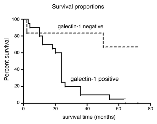
Figure 3. Survival analysis of galectin-1 immunostaining and patient survival in validation cohort 2 (Cleveland Clinic cohort). Patients with negative galectin-1 staining (IHC score 0) had significantly longer survival than the patients with positive galectin-1 staining (IHC score ≥ 1+) with hazard ratio 4.9 (p = 0.002).

Table 2. Galectin-1 staining in Pancreatic ductal adenocarcinoma TMA

Figure 4. Representative galectin-1 staining of pancreas tissue microarrays. All ductal epithelial cells and neoplastic cells were negative for galectin-1 staining. The positive staining was detected in the stromal cells for galectin-1 positive specimens. A. Normal pancreas (no staining); B. Normal pancreas adjacent to PDAC (1+); C. Primary benign chronic pancreatitis (1+); D. PanIN 2 (2+); E. PanIN3 (2+); F. PDAC (3+).
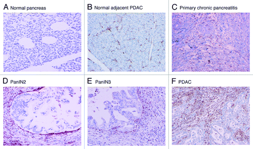
Figure 4. Representative galectin-1 staining of pancreas tissue microarrays. All ductal epithelial cells and neoplastic cells were negative for galectin-1 staining. The positive staining was detected in the stromal cells for galectin-1 positive specimens. A. Normal pancreas (no staining); B. Normal pancreas adjacent to PDAC (1+); C. Primary benign chronic pancreatitis (1+); D. PanIN 2 (2+); E. PanIN3 (2+); F. PDAC (3+).
Supplemental material

Additional material

Download Zip (81 KB)

Reprints and Corporate Permissions

Please note: Selecting permissions does not provide access to the full text of the article, please see our help page How do I view content?

To request a reprint or corporate permissions for this article, please click on the relevant link below:

Academic Permissions

Please note: Selecting permissions does not provide access to the full text of the article, please see our help page How do I view content?

Obtain permissions instantly via Rightslink by clicking on the button below:

If you are unable to obtain permissions via Rightslink, please complete and submit this Permissions form. For more information, please visit our Permissions help page.