1,068
Views
11
CrossRef citations to date
0
Altmetric
Research Paper

Interferon-gamma enhances radiation-induced cell death via downregulation of Chk1

, &
Pages 1018-1025 | Received 16 Feb 2012, Accepted 04 Jun 2012, Published online: 24 Jul 2012

Figures & data

Figure 1. IFNγ treatment induced S-phase delay and ATR/Chk1 phosphorylation. (A) HeLa cells were treated with the indicated concentrations of IFNγ for 4 d, and cell proliferation was measured with the MTT assay. (B) Cells were treated with 200 U/ml IFNγ for 2 d, and cell cycle profiles were obtained by flow cytometry. Values are means ± standard deviation (SD) of three independent experiments. (C) IFNγ treatment activates Chk1 via ATR. HeLa cells were treated with 200 U/ml IFNγ up to 24 h, and protein expression was determined by western blot analysis. β-actin was used as a loading control. (D) Cells were treated with 0, 100, 200, or 400 U/ml of IFNγ for 12 h, and ATM, ATR and Chk1 phosphorylation was determined by western blot analysis.
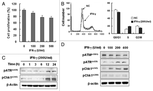
Figure 1. IFNγ treatment induced S-phase delay and ATR/Chk1 phosphorylation. (A) HeLa cells were treated with the indicated concentrations of IFNγ for 4 d, and cell proliferation was measured with the MTT assay. (B) Cells were treated with 200 U/ml IFNγ for 2 d, and cell cycle profiles were obtained by flow cytometry. Values are means ± standard deviation (SD) of three independent experiments. (C) IFNγ treatment activates Chk1 via ATR. HeLa cells were treated with 200 U/ml IFNγ up to 24 h, and protein expression was determined by western blot analysis. β-actin was used as a loading control. (D) Cells were treated with 0, 100, 200, or 400 U/ml of IFNγ for 12 h, and ATM, ATR and Chk1 phosphorylation was determined by western blot analysis.

Figure 2. IFNγ treatment induces instability of Chk1 protein. (A) HeLa cells were treated with 200 U/ml IFNγ up to 24 h, and Chk1 expression was measured by western blot analysis. (B) Cells were treated with 0, 100, 200, or 400 U/ml of IFNγ for 12 h, and Chk1 protein levels were determined by western blot analysis. (C) Cells were treated with 200 U/ml IFNγ for 0, 1, 3, 6, or 12 h, and Chk1 mRNA levels were determined by RT-PCR. (D) Cells were treated with 100 μg/ml cyclohexamide (CHX) and cultured for the indicated times after 12 h treatment with 200 U/ml IFNγ. Chk1 protein levels were determined by western blot analysis. β-actin was used as a loading control. The figure shows representative data from three independent experiments.
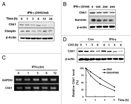
Figure 2. IFNγ treatment induces instability of Chk1 protein. (A) HeLa cells were treated with 200 U/ml IFNγ up to 24 h, and Chk1 expression was measured by western blot analysis. (B) Cells were treated with 0, 100, 200, or 400 U/ml of IFNγ for 12 h, and Chk1 protein levels were determined by western blot analysis. (C) Cells were treated with 200 U/ml IFNγ for 0, 1, 3, 6, or 12 h, and Chk1 mRNA levels were determined by RT-PCR. (D) Cells were treated with 100 μg/ml cyclohexamide (CHX) and cultured for the indicated times after 12 h treatment with 200 U/ml IFNγ. Chk1 protein levels were determined by western blot analysis. β-actin was used as a loading control. The figure shows representative data from three independent experiments.

Figure 3. IFNγ treatment increases radiosensitivity. (A) HeLa cells were treated with 200 U/ml IFNγ for 12 h before irradiation (5Gy), and the levels of indicated proteins were determined by western blotting. (B) Cleavage of PARP and Caspase3 was measured after treatment with IFNγ and IR by western blotting. (C) Cells were treated with 200 U/ml IFNγ and IR for 2 d and stained with Annexin V-FITC and propidium iodide (PI). The fluorescence intensity of Annexin V-FITC was quantified by flow cytometry. (D) The percentage of the population undergoing apoptosis was calculated by flow cytometry with Annexin V-FITC and PI. Values are means ± SD of three independent experiments.
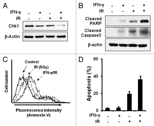
Figure 3. IFNγ treatment increases radiosensitivity. (A) HeLa cells were treated with 200 U/ml IFNγ for 12 h before irradiation (5Gy), and the levels of indicated proteins were determined by western blotting. (B) Cleavage of PARP and Caspase3 was measured after treatment with IFNγ and IR by western blotting. (C) Cells were treated with 200 U/ml IFNγ and IR for 2 d and stained with Annexin V-FITC and propidium iodide (PI). The fluorescence intensity of Annexin V-FITC was quantified by flow cytometry. (D) The percentage of the population undergoing apoptosis was calculated by flow cytometry with Annexin V-FITC and PI. Values are means ± SD of three independent experiments.

Figure 4. XAF1 is involved in IFNγ-induced Chk1 downregulation. (A) HeLa cells were treated with 200 U/ml IFNγ for the indicated times, and IRF-1 and XAF1 protein levels were measured by western blot analysis. (B) HeLa cells were transfected with IRF-1 siRNA and treated with 200 U/ml IFNγ for 12 h. The levels of IRF1, XAF1 and Chk1 were analyzed by western blotting. (C) Cells were transfected with XAF1, or a control vector and incubated for 36 h. The level of Chk1 was analyzed by western blotting. (D) Cells were transfected with XAF1 siRNA and treated with 200 U/ml IFNγ for 12 h. The level of Chk1 was analyzed by western blotting.
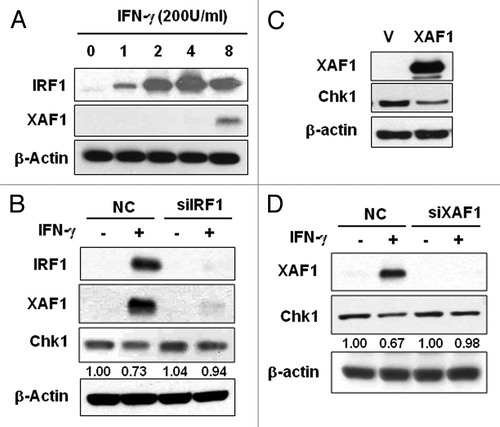
Figure 4. XAF1 is involved in IFNγ-induced Chk1 downregulation. (A) HeLa cells were treated with 200 U/ml IFNγ for the indicated times, and IRF-1 and XAF1 protein levels were measured by western blot analysis. (B) HeLa cells were transfected with IRF-1 siRNA and treated with 200 U/ml IFNγ for 12 h. The levels of IRF1, XAF1 and Chk1 were analyzed by western blotting. (C) Cells were transfected with XAF1, or a control vector and incubated for 36 h. The level of Chk1 was analyzed by western blotting. (D) Cells were transfected with XAF1 siRNA and treated with 200 U/ml IFNγ for 12 h. The level of Chk1 was analyzed by western blotting.

Figure 5. XAF1 is involved in IFNγ-induced radiosensitization. (A) HeLa cells were transfected with XAF1 siRNA and treated with 200 U/ml IFNγ for 12 h before irradiation (5Gy). The levels of indicated proteins were determined by western blotting. (B) Cell viability of the cells after irradiation was measured with MTT assay. Results were expressed as a percent cell survival compared with the control. Values are means ± SD of three experiments. *p < 0.05 vs. control. (C) Clonogenic cell survival assay. HeLa cells were transfected with control vector (V), XAF1 expression vector (XAF1), Chk1 expression vector (Chk1) or Chk1 siRNA (siChk1) and incubated for 36 h. Control for Chk1 siRNA is indicated as C. After each of cells reseeded and incubated for 24 h, cells were irradiated with 0, 1, 2 and 4 Gy of IR. Number of colonies were counted and expressed in comparison to 0 Gy control sample. Values are means ± SD of three independent experiments.
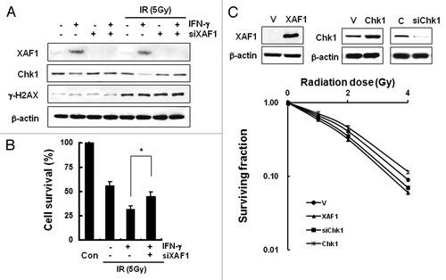
Figure 5. XAF1 is involved in IFNγ-induced radiosensitization. (A) HeLa cells were transfected with XAF1 siRNA and treated with 200 U/ml IFNγ for 12 h before irradiation (5Gy). The levels of indicated proteins were determined by western blotting. (B) Cell viability of the cells after irradiation was measured with MTT assay. Results were expressed as a percent cell survival compared with the control. Values are means ± SD of three experiments. *p < 0.05 vs. control. (C) Clonogenic cell survival assay. HeLa cells were transfected with control vector (V), XAF1 expression vector (XAF1), Chk1 expression vector (Chk1) or Chk1 siRNA (siChk1) and incubated for 36 h. Control for Chk1 siRNA is indicated as C. After each of cells reseeded and incubated for 24 h, cells were irradiated with 0, 1, 2 and 4 Gy of IR. Number of colonies were counted and expressed in comparison to 0 Gy control sample. Values are means ± SD of three independent experiments.

Table 1. Cell death measurement of lung cancer cell lines following combined treatment with IFNγ and IR

Reprints and Corporate Permissions

Please note: Selecting permissions does not provide access to the full text of the article, please see our help page How do I view content?

To request a reprint or corporate permissions for this article, please click on the relevant link below:

Academic Permissions

Please note: Selecting permissions does not provide access to the full text of the article, please see our help page How do I view content?

Obtain permissions instantly via Rightslink by clicking on the button below:

If you are unable to obtain permissions via Rightslink, please complete and submit this Permissions form. For more information, please visit our Permissions help page.