Figures & data
Table 1. Clinic-pathological characteristic of study population
Figure 1. EGFR expression in normal peri-tumor oral epithelium as evaluated by immunohistochemistry and in representative cases of OSCC’swith amplified EGFR. (A) EGFR shows a high expression limited to the basal proliferative layer, whereas spinous epithelial layer demonstrated faint-intermediate expression at membrane malpighian bridges ( LSAB-HRP, original magnification x63, inset original magnification x100). (B) Photos A and B show two different cases with strong EGFR expression. Amplified cases showed very strong circumferential staining of the membrane that appeared remarkable thickened; in addition, cytoplasms were stained (A1 and B1) and in some fields (A1) the nuclei appeared positive (LSAB-HRP, nuclear counterstaining with haematoxylin; Ventana pre-diluted Ab not specific for activating phosphorylations).
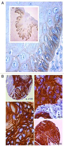
Figure 2. Phosphorylated EGFR-p-tyr1068 and p-tyr845 in oral cancer as evaluated by TMA-based immunohistochemistry. (A) A TMA core of positive p-EGFR-tyr 1068 is showed in (A), at larger magnification in (B), demonstrating strong cytoplasmic expression of the activated receptor (LSAB-HRP, nuclear counterstaining with haematoxylin). (B) Phosphorylation of EGFR on Tyrosine 845 in oral cancer as evaluated by TMA-based IHC. A, A1, A2 show high cytoplasmic expression of p-845 EGFR in a representative case of OSCC with vascular invasion. B, B1, B2 show p-845 EGFR in a case of OSCC with poor differentiation (A, A1, A2, B, B1, B2: IHC- LSAB-HRP, nuclear counterstaining with haematoxylin; phosphorylated tyr845 EGFR Ab).
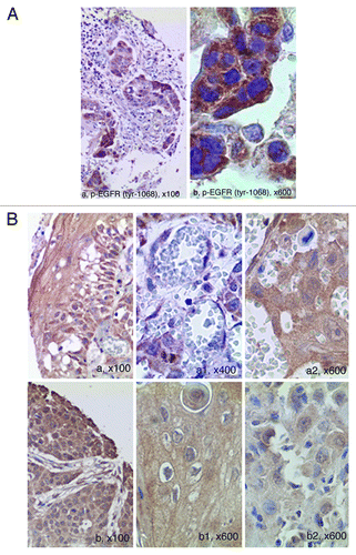
Table 2. Clinic-pathological characteristics of pEGFR Tyr 845/1068 positive and negative OSCCs
Figure 3. Western blotting of representative normal epithelia, EGFR-trisomic and EGFR-amplified OSCC’s using phosphorylated Tyr 845 EGFR antibody. Both trisomic and amplified OSCC’s show EGFR phosphorylation on both Tyr sites, whereas normal epithelia are negative. For further details see “Materials and Methods” section.
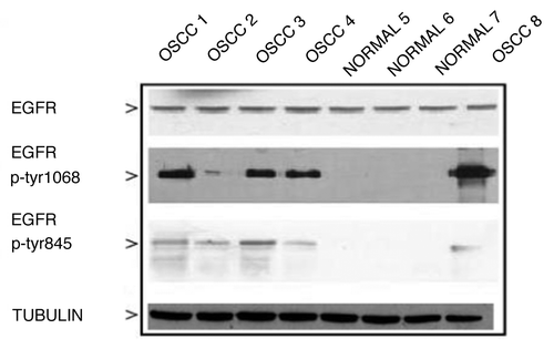
Figure 4. FISH analysis for the detection of EGFR amplification. (A) An OSCC showing euploid chromosome 7 and not amplified EGFR; (B) a case of OSCC showing triploid chromosome 7 and three copies of EGFR; (C) a case of OSCC demonstrating aneuploidy at chromosome 7 and multiple copies of EGFR (polysomic not amplified EGFR); (D) a case of OSCC showing euploid chromosome 7 and multiple copies of EGFR visualized as nuclear clusters (amplified EGFR gene) [FISH: LSI EGFR Dual-Color Probe-Hyb Set, LSI EGFR Spectrum Orange/Cep-7 Spectrum Green; DAPI II (4,6-diamino-2-phenyindole-2-hydrochloride) was used for chromatin counterstaining; a ratio of LSI EGFR Spectrum Orange/Cep-7 Spectrum Green >2 has been considered as amplified; for further details see Materials and Methods].
![Figure 4. FISH analysis for the detection of EGFR amplification. (A) An OSCC showing euploid chromosome 7 and not amplified EGFR; (B) a case of OSCC showing triploid chromosome 7 and three copies of EGFR; (C) a case of OSCC demonstrating aneuploidy at chromosome 7 and multiple copies of EGFR (polysomic not amplified EGFR); (D) a case of OSCC showing euploid chromosome 7 and multiple copies of EGFR visualized as nuclear clusters (amplified EGFR gene) [FISH: LSI EGFR Dual-Color Probe-Hyb Set, LSI EGFR Spectrum Orange/Cep-7 Spectrum Green; DAPI II (4,6-diamino-2-phenyindole-2-hydrochloride) was used for chromatin counterstaining; a ratio of LSI EGFR Spectrum Orange/Cep-7 Spectrum Green >2 has been considered as amplified; for further details see Materials and Methods].](/cms/asset/4581b544-a85e-4a69-b103-0436ad1a45d4/kcbt_a_10920991_f0004.gif)
Table 3. Frequencies of FISH amplification according to pathological correlations
Figure 5. Kaplan Meier curvesof patients positive for p-Tyr 845 and 1068EGFR stratified for gender. Survival curves showing percentage survival according to sex and EGFR-p-Tyr 845 (A, B), and EGFR-p-Tyr 1068 (C, D). Interestingly, inside the group of females affected by OSCC the EGFR-p-Tyr 845 negative subgroup showed a trend for a better survival if compared to the EGFR-p-Tyr 845 positive subgroup (for details, see the text).
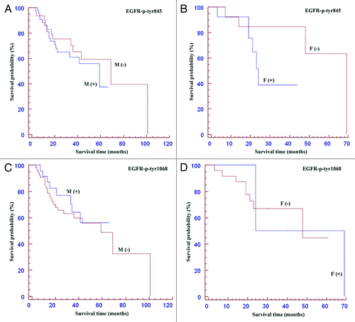
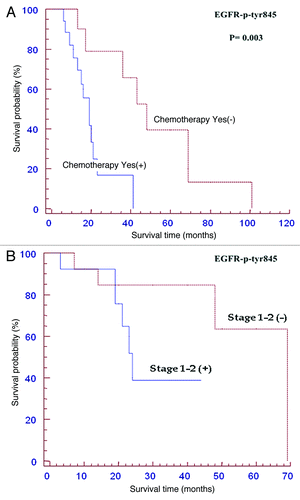
Figure 6. Kaplan Meier curves of patients positive forp-Tyr-845 EGFR expression subjected to adjuvant chemotherapy (A) or with early stage tumours (B). Among patients receiving adjuvant chemotherapy p-Tyr 845 negative cases had a better survival (p<0.05). Among T1-T2 tumors p-Tyr 845 negative cases had a better survival (p<0.05). See the text for details.