Figures & data
Figure 6. Nutlin-3 protects only p53 wild-type cells from S phase-specific drug combinations. (A) Cells were incubated with 0–10 ng/ml SN38 alone or in combination with 10 µM KU55933 (ATM inhibitor) for 24 h. (B) Cells were incubated with 0–10 mM hydroxyurea alone or in combination with 1 µM MK-8776 (Chk1 inhibitor) for 24 h. Cells were also pretreated with 10 µM Nutlin-3 for 24 h, and this was left on during incubation with the other drugs. Drugs were removed 24 h later and cells were collected 5–7 d later when the untreated or Nutlin-3 only treated cells reached 70–80% confluency. The error bars represent SE of at least two independent experiments.
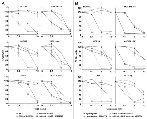
Figure 1. p53 and p21waf1 protein levels induced by SN38 and Nutlin-3. (A) Cells were incubated with 10 ng/ml SN38 or 10 µM Nutlin-3 for 24 h. Protein levels were assessed by western blotting. (B) The ratio of p21waf1 to p53 protein levels were compared with that of MCF10A after 24 h of SN38.
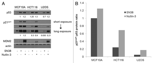
Figure 2A–C. Protein kinetics during and following treatment with Nutlin-3. (A) MCF10A, (B) HCT116 and (C) U2OS were incubated with 10 µM Nutlin from 0–24 h. The drug was removed and cells were incubated for an additional 24 h in fresh medium. Cells were harvested at the indicated times and assayed for proteins using western blotting.
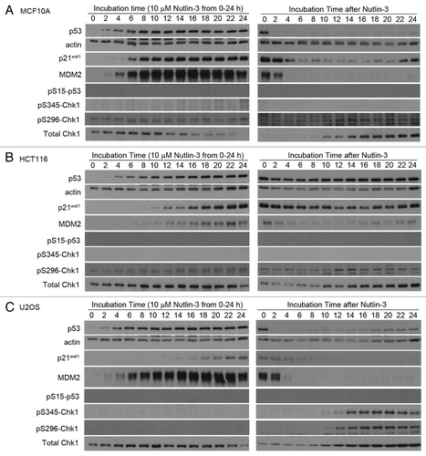
Figure 2D and E. Protein kinetics during and following treatment with Nutlin-3. (D) p53 and E. p21waf1 protein levels were quantified by densitometry of multiple exposures of western blots. The left panels show protein induction compared with the untreated control of each cell line. The right panels show protein induction compared with untreated MCF10A cells.
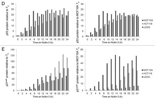
Figure 3. p21waf1 mRNA kinetics following Nutlin-3 treatment. (A) Cells were incubated with 10 µM Nutlin-3 from 0–24 h. The drug was removed and cells were incubated for an additional 24 h in fresh medium. Cells were harvested at the indicated times and assayed for p21waf1 mRNA levels using RT-qPCR. (B) Cells were incubated with 10 ng/ml SN38 or 10 µM Nutlin-3 for 24 h, harvested and assayed for p21waf1 mRNA levels using RT-qPCR. The error bars represent SE of at least three independent experiments.
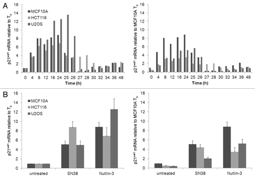
Figure 4. Higher Nutlin-3-induced p21waf1 protein levels is due to longer p21waf1 protein half-life. Cells were incubated with 10 µM Nutlin-3 for 24 h and then incubated with the protein synthesis inhibitor cycloheximide (10 µg/ml) for the indicated times without removing nutlin-3. The densitometric scans of the western blots are graphed on the right. The half-lives were calculated from three such independent experiments and are presented as the means ± SE. The half-lives for SN38 are derived from our previous paper (11).
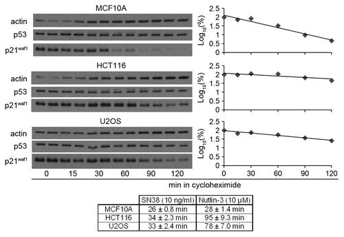
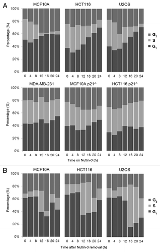
Figure 5. Nutlin-3-induced p21waf1 protein leads to reversible G1/G2 arrest. (A) p53 wild-type cells (top panel) and p53- and p21waf1-defective cells (bottom panel) were incubated with 10 µM Nutlin-3 for 0–24 h. Cells were harvested and assayed for DNA content by flow cytometry. (B) After 24 h of 10 µM Nutlin-3, the drug was removed and cells were incubated for an additional 24 h in fresh medium before assaying by flow cytometry.