Figures & data
Figure 1. Greater suppression of HIF-1α with BEZ235 than LY294002 or rapamycin. (A and B) Cells were treated with BEZ235, LY294002 or rapamycin for 3 h, and thereafter cells were exposed to 0.5% oxygen (lanes 6-10) for 3 h before cells were harvested. (C and D) Cells were treated with 100 nM BEZ235 for 24 h, followed by addition of MG132 for 1, 2, 3 and 4 h. After MG132 treatment, protein was harvested. For panels A–D, proteins samples were analyzed by SDS-PAGE and immunoblotting using antibodies as indicated, including HIF-1α and β-actin (loading control).
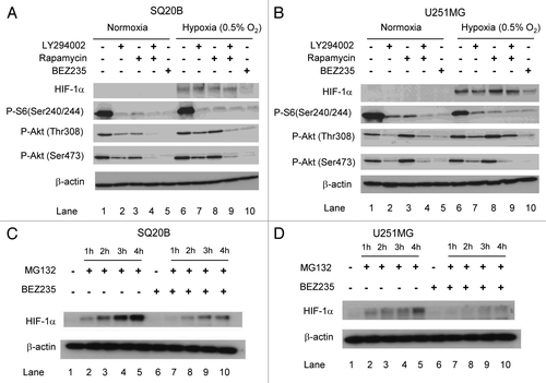
Figure 2. BEZ235 treatment results in decreased global protein translation. (A) SQ20B or U251MG cells were treated with BEZ235 for 24 h. 30 minutes before the end of treatment, cells were labeled with [35S]methionine (50 µCi/ml). At completion of assay, cells were lysed and assessed for total protein content and radioactivity. Results are represented as the counts per minute (cpm) normalized to total protein concentration in the drug-treated cells compared to the untreated control cells. (B) The effect of PI3K/Akt/mTOR inhibition on cap dependent translation. The figure shows the interactions between eIF4E, 4E-BP1 and eIF4G. (C) SQ20B or U251MG cells were treated with drugs as indicated for 3 h. Whole cell extracts were prepared and eIF4E and its associated proteins purified by 7-methyl-GTP-Sepharose affinity chromatography. (D) SQ20B or U251MG cells were treated with 4EGI1 or BEZ235 for 24 h. After the drug treatment cells were exposed to hypoxia for 3 h. Thereafter, protein was harvested. For panels C and D, protein samples were analyzed by SDS-PAGE and immunoblotting for proteins using antibodies as shown.
![Figure 2. BEZ235 treatment results in decreased global protein translation. (A) SQ20B or U251MG cells were treated with BEZ235 for 24 h. 30 minutes before the end of treatment, cells were labeled with [35S]methionine (50 µCi/ml). At completion of assay, cells were lysed and assessed for total protein content and radioactivity. Results are represented as the counts per minute (cpm) normalized to total protein concentration in the drug-treated cells compared to the untreated control cells. (B) The effect of PI3K/Akt/mTOR inhibition on cap dependent translation. The figure shows the interactions between eIF4E, 4E-BP1 and eIF4G. (C) SQ20B or U251MG cells were treated with drugs as indicated for 3 h. Whole cell extracts were prepared and eIF4E and its associated proteins purified by 7-methyl-GTP-Sepharose affinity chromatography. (D) SQ20B or U251MG cells were treated with 4EGI1 or BEZ235 for 24 h. After the drug treatment cells were exposed to hypoxia for 3 h. Thereafter, protein was harvested. For panels C and D, protein samples were analyzed by SDS-PAGE and immunoblotting for proteins using antibodies as shown.](/cms/asset/2303ddfb-5300-43f1-8dde-38f0b9df3811/kcbt_a_10921144_f0002.gif)
Figure 3. Effects of BEZ235 on survival under hypoxia. (A) SQ20B cells were exposed to 0.5% O2 for 24 h, and then treated with BEZ235 for 24 h. The cell viability was measured by the MTT assay. (B) SQ20B cells were exposed to 0.5% O2 for 24 h, and then treated with BEZ235 for 24 h. After 10 days the colonies were stained and counted. The graph underneath shows the surviving fraction ratios under hypoxia and normoxia (plating efficiency in the presence of BEZ235 divided by plating efficiency in the controls without the drug). (C) SQ20B cells were exposed to 0.5% O2 for 24 h, then treated with BEZ235 for 1, 3, 6, 16 and 24 h prior to protein harvesting. Proteins were analyzed by SDS-PAGE and immunoblotting with antibodies as shown. (D) SQ20B cells were exposed to 0.5% O2 for 24 h, and then treated with BEZ235 for 24 h. Cell death detection ELISA was used to quantitate the cytoplasmic oligonucleosomes as a measure of apoptosis.
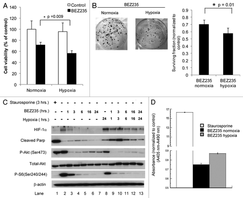
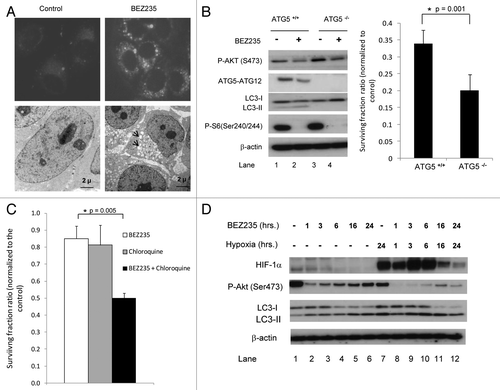
Figure 4. Effects of BEZ235 on autophagy. (A) Upper panel. SQ20B cells were stably transfected with GFP-LC3. The cells were then treated with or without BEZ235 for 24 h and examined by fluorescence microscopy. Lower panel. SQ20B cells were treated with BEZ235 for 24 h, fixed and analyzed by electron microscopy. There were numerous vacuoles in the cytoplasm of the drug treated cells (arrows). (B) ATG5 wild-type (ATG5+/+) and knockout (ATG5-/-) MEFs were treated with BEZ235 for 24 h. Left panel. After drug treatment cells were harvested for protein. Right panel. Cells were refed with drug free DMEM and after 10 days the colonies were stained and counted. The graph shows the surviving fraction ratios in ATG5+/+ and ATG5-/- cells (plating efficiency in the presence of BEZ235 divided by plating efficiency in the controls without the drug). (C) SQ20B cells were treated either with BEZ235 or chloroquine or a combination of these drugs for 6 h. After 10 days the colonies were stained and counted. Graph shows the surviving fraction ratios in SQ20B cells (plating efficiency in the presence of drug(s) divided by plating efficiency in the controls without drug). (D) SQ20B cells were exposed to 0.5% O2 for 24 h, then treated with BEZ235 for 1, 3, 6, 16 and 24 h prior to protein harvesting. Proteins were analyzed by SDS-PAGE and immunoblotting using antibodies as shown.