Figures & data
Table 1. IC50 values of chemotherapeutic agents. IC50 values (µM) were determined based on cellular protein remaining after 72 h incubation
Figure 1. Mitochondrial status. (A) western blots showing expression of Bcl-2, Bcl-xL and ABCG2 in A2780 parental (AP) and cisplatin-resistant (AR) cells, SKOV-3 parental (P), cisplatin-resistant (R) and SKOV-3-ρ0 cells (ρ0). GAPDH served as loading control on both blots. (B) Cellular mitochondrial mtDNA was quantitated as the ratio of mitochondrial DNA (mtDNA) to nuclear β-actin DNA, and was assessed for three different mtDNA genes (12S, Cox-I and ND4L). P: SKOV-3 parental cells; R: SKOV-3-R; ρ0: SKOV-3-ρ0. Values from the SKOV-3-ρ0 cells were too low to be depicted clearly. Data are from three separate DNA preparations, each analyzed in duplicates using qRT-PCR. The increase in Cox-I: β-actin ratio in SKOV-3-R was statistically significant (p = 0.025) as well as the decreased ratio for all three genes in SKOV-3-ρ0 (all p < 0.001). Error bars represent SEM (C) Cellular mitochondrial mass was assessed using MitoTracker Green FM dye. The bars represent flow cytometry results from three separate quantitations with triplicate samples in each. The increase in mitochondrial mass in SKOV-3-R was statistically significant (p = 0.011). Error bars represent SEM (D-E) western blots showing expression of voltage-dependent anion channel (VDAC) and cytochrome c (cyto c) (D) and hexokinase-II (HK-II) (E) in the three cell lines. Values underneath each blot are densitometry-based estimations of fold increase in signal compared with SKOV-3 parental cells, and corrected for loading based on β-tubulin or GAPDH signal.
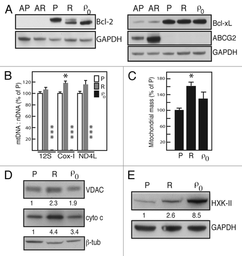
Figure 2. EMT and motility. (A) Phase contrast microscope (40x magnification) photomicrographs of SKOV-3 (P), SKOV-3-R (R) and SKOV-3-ρ0 (ρ0) cells. (B) western blots showing expression of E-cadherin (E-cadh), vimentin (vim) and Twist in the same cell lines. GAPDH served as loading control. (C) Immunofluorescence photomicrographs (60x magnification) showing levels and nuclear location of transcriptional repressors Twist and Snail, respectively, in SKOV-3 parental (P) and resistant (R) cells. Red: Twist or Snail; blue: DAPI nuclear stain. (D) Representative tracings of SKOV-3 (P) and SKOV-3-R (R) cells over 18 h from the automated assessment of motility toward a chemoattractant (serum-containing medium), using xCELLigence® equipment. Error bars represent standard deviation in quadruplicates. The experiment was performed three times with similar results.
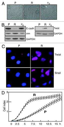
Figure 3. Stem cell features of SKOV-3-R cells. (A) western blots showing protein levels of ovarian cancer stem cell (tumor-initiating cell, TIC) markers CD117, CD44 and ALDH1. GAPDH served as loading control. P: SKOV-3 parental cells; R: SKOV-3-R; ρ0: SKOV-3-ρ0. (B) Immunofluorescence photomicrographs showing co-staining for CD117 and E-cadherin in SKOV-3 (P) and SKOV-3-R (R) cells. Green: CD117; Red: E-cadherin; Blue: DAPI nuclear stain. (C) Based on three separate experiments, assessment of percent cells with the indicated presence or absence of E-cadherin and CD117, respectively, in SKOV-3 parental (black) and SKOV-3-R cells (gray). p = 0.039 and p < 0.001, respectively. (D) Representative photomicrographs (20x magnification) of each cell line after 19 d in stem cell medium. Each cell line was seeded in stem cell medium and inspected daily. The SKOV-3 parental and SKOV-3-ρ0 cells did not form spheres and instead gradually rounded up and died. By contrast, after 3 d SKOV-3-R cells began to form spheres that were viable, i.e., could then be dispersed, replated and grown both in stem cell medium as spheres, and in standard medium as adherent cells. SCM: stem cell medium; S: standard medium. The experiment was repeated with identical results.
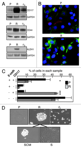
Table 2. Targeting the multiresistant SKOV3-R cells.
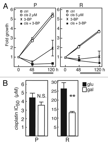
Figure 4. Targeting multiresistant SKOV-3-R cells. (A) Combination treatments of cisplatin (cis; 2 and 5 µM) and 3-bromopyruvate (3-BP; 30 µM). SKOV-3 (P; left) and SKOV-3-R (R; right) cells were treated with drugs, singly and in combination, for 48 h, whereafter all supernatant media were replaced with fresh medium without drugs and cells were allowed to regrow for another 72 h (in total 120 h). The gray bar underneath each graph indicates this drug-free incubation. Cellular protein at each time point was assessed using the SRB assay, and data are expressed as fold increase from t = 0. Data represent averages from three separate experiments with quadruplicate samples in each. SEM error bars were too small to be visualized, except where shown. For SKOV-3-R cells, data for 5 µM cisplatin are shown, but 2 µM yielded nearly identical results. Thus, the 120 h endpoint values were 0.38 for 5 µM + 3-BP, and 0.60 for 2 µM + 3-BP. (B) SKOV-3 parental (P; left) and SKOV-3-R (R; right) cells were cultured in standard medium containing glucose (glu; black bars) and in galactose medium without glucose (gal; white bars), respectively. The IC50:s of cisplatin, based on six concentrations, were determined after 72 h incubation in each type of medium. Results are based on averages from at least four separate experiments with each concentration in quadruplicate. Error bars represent SEM. N.S., not significant, p = 0.187. **p = 0.004.