Figures & data
Figure 1. Effects of dasatinib and sunitinib on Cav-1 expression and secretion and on TK signaling in PC-3 cells. Dasatinib (A) and sunitinib (B) treatment of PC-3 cells resulted in a dose-dependent decrease in phosphorylation of PDGFRβ, VEGFR2, Akt and Cav-1. Dasatinib but not sunitinib also reduced the phosphorylation of FAK and Src. Both dasatinib and sunitinib dose-dependently reduced the expression and secretion of Cav-1.
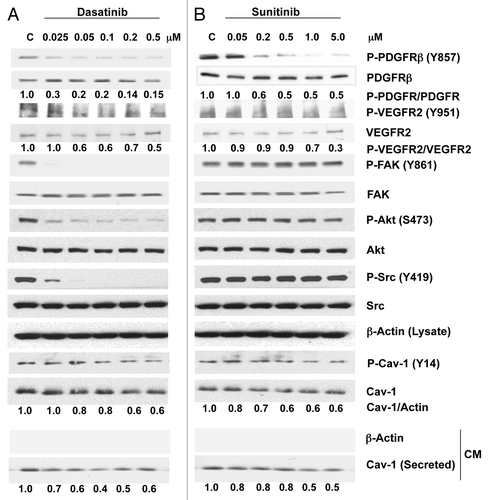
Figure 2. Effects of dasatinib and sunitinib on Cav-1 expression and secretion and on TK signaling in DU145 cells. Dasatinib (A) and sunitinib (B) treatment of DU145 cells resulted in a dose-dependent decrease in phosphorylation of PDGFRβ, VEGFR2, Src and Cav-1. Dasatinib, but not sunitinib, also reduced the phosphorylation of FAK and Akt. Both dasatinib and sunitinib dose-dependently reduced significantly the secretion of Cav-1.
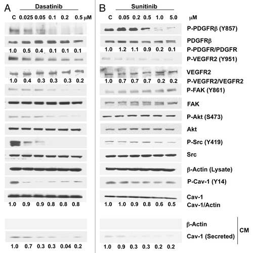
Figure 3. Effects of dasatinib and sunitinib on PC-3 and DU145 cellular proliferation. Dasatinib (Dasa) inhibited proliferation of PC-3 (A) cells and DU145 cells (B) at doses ranging from 0.05–5.0 μM Sunitinib (Suni) inhibited proliferation of PC-3 cells (C) and DU145 cells (D) at doses ranging from 0.2–20 μM. Data are plotted as means ± SD of experiments repeated in triplicate.*, p < 0.05; **, p < 0.0001 by Student’s t-test compared with control group.
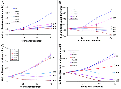
Figure 4. Antitumor activity of dasatinib (Dasa) in combination with anti-Cav-1 antibody (Cav-1 Ab) on PC-3 xenografts in nude mice. (A) Dasatinib alone and anti-Cav-1 antibody alone significantly reduced the tumor volume of PC-3 cells growing as xenografts compared with those of vehicle- and IgG-treated controls (p = 0.002 and p = 0.02, respectively). Anti-Cav-1 antibody treatment also enhanced the efficacy of dasatinib. Each data point represents the mean tumor volume in each group containing 9–16 mice (B), dasatinib alone and anti–Cav-1 antibody alone significantly reduced the tumor wet weight in nude mice bearing PC-3 tumor xenografts compared with the weights in vehicle- and IgG-treated controls (p = 0.0072 and p = 0.0307, respectively). Data represents the mean tumor weight ± SEM in each group containing 9–16 mice.
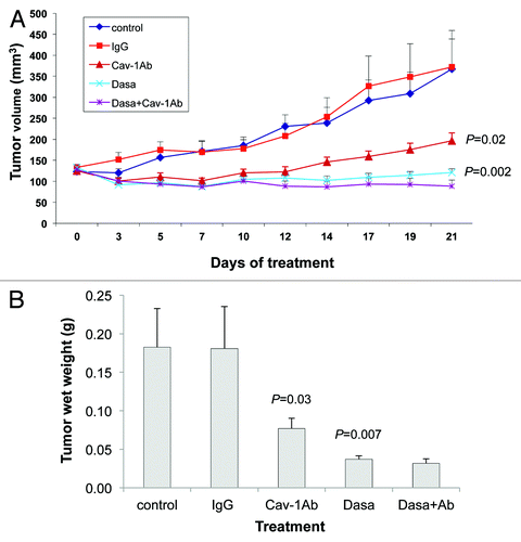
Figure 5. Antitumor activity of sunitinib (Suni) alone and in combination with anti–Cav-1 antibody (Cav-1Ab) on DU145 xenografts in nude mice. (A) Sunitinib alone and anti-Cav-1 antibody alone significantly reduced the tumor volume of DU145 cells growing as xenografts compared with those of vehicle- and IgG-treated controls (p = 0.0015 and p = 0.0377, respectively). Anti-Cav-1 antibody treatment also enhanced the efficacy of sunitinib. Each data point represents the mean tumor volume in each group containing 7–10 mice. (B) Sunitinib alone and anti-Cav-1 antibody alone significantly reduced the tumor wet weight in nude mice bearing DU145 tumor xenografts compared with those in vehicle- and IgG-treated control mice (p = 0.0004 and p = 0.0016, respectively). Data represents the mean tumor weight ± SEM in each group containing 7–10 mice.
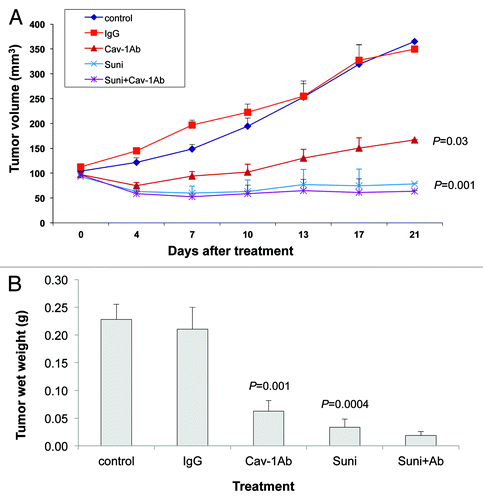
Figure 6. Dasatinib and sunitinib suppress RTK signaling by downregulation of Cav-1. (A) Serum Cav-1 concentrations in mice treated with dasatinib were significantly reduced compared with those in the control group (p = 0.027). (B) Serum Cav-1 concentration in mice treated with sunitinib were reduced compared with that in the control group, although the differences did not achieve statistical significance (p = 0.0871). (C) Cav-1 knockdown in PC-3 cells treated with dasatinib or sunitinib caused greater reduction in the phosphorylation of PDGFRβ and VEGFR2 than control siRNA, and suppressed cellular and secreted PDGF-B and VEGF-A [secreted VEGF-A were reduced to undetectable levels (un) in response to Cavsi alone and Cavsi dasatinib or sunitinib treatment]. (D) Dasatinib and sunitinib inhibit PDGFRβ and VEGFR2 signaling by downregulation of Cav-1 expression and secretion, in turn, leads to suppression of expression and secretion of PDGF-B and VEGF-A. Data in (A and B) represent mean serum Cav-1 + SD in each group.
![Figure 6. Dasatinib and sunitinib suppress RTK signaling by downregulation of Cav-1. (A) Serum Cav-1 concentrations in mice treated with dasatinib were significantly reduced compared with those in the control group (p = 0.027). (B) Serum Cav-1 concentration in mice treated with sunitinib were reduced compared with that in the control group, although the differences did not achieve statistical significance (p = 0.0871). (C) Cav-1 knockdown in PC-3 cells treated with dasatinib or sunitinib caused greater reduction in the phosphorylation of PDGFRβ and VEGFR2 than control siRNA, and suppressed cellular and secreted PDGF-B and VEGF-A [secreted VEGF-A were reduced to undetectable levels (un) in response to Cavsi alone and Cavsi dasatinib or sunitinib treatment]. (D) Dasatinib and sunitinib inhibit PDGFRβ and VEGFR2 signaling by downregulation of Cav-1 expression and secretion, in turn, leads to suppression of expression and secretion of PDGF-B and VEGF-A. Data in (A and B) represent mean serum Cav-1 + SD in each group.](/cms/asset/ed576f94-d4b0-406a-82f3-a8c059ca217d/kcbt_a_10922633_f0006.gif)