Figures & data
Figure 1. Low fluorescence of imaging diet (ID) in the NIR wavelength and weight loss of mice subjected to a DSS-induced IBD. (A) Imaging diet (ID) provides better noise-to-signal compared to standard diet (SD) in the NIR wavelength of light compared to "green fluorescent" wavelength of light (515 nm). B/F-bright field. (B) Weight loss of female and male C57BL/6J mice subjected to DSS (2% w/v) in the drinking water. *P < 0.05, the Student t test.
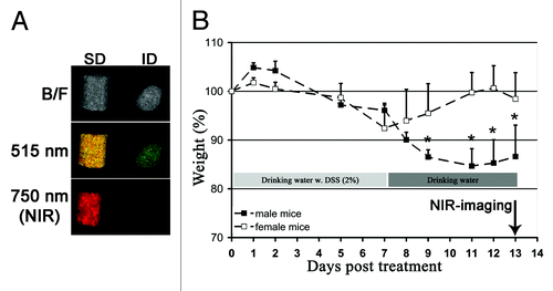
Figure 2. Whole-body non-invasive NIR optical imaging confirms DSS-induced IBD in mice injected with ProSense680. (A) Statistically significant differences in body weight between female and male C57BL/6J mice subjected DSS-induced IBD and/or non-invasive NIR-optical imaging (P < 0.0001, 2-way ANOVA). (B) NIR optical imaging (“non-invasive” and concomitant with necropsy) of female (f) and male (m) mice injected with ProSense680 reveals increased unmixed signals from male mice subjected to DSS (2% w/v) in the drinking water consistent with increased epithelial atrophy and inflammation in males compared to females as detected by H/E (C). Representative images are shown. (D) Ex vivo NIR imaging of “swiss-rolled” small intestines and colons from female and male mice with DSS-induced IBD. Duo, duodenum; jej, jejunum; ile,ileum; dc, descending colon; tc, transcending colon; ccc, cecum.
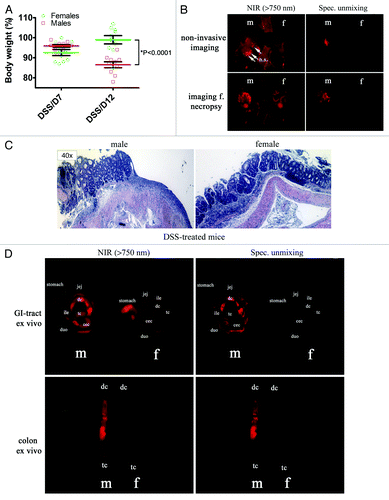
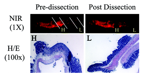
Figure 3. NIR-imaging identifies inflammatory hot spots of the colon of male mice with DSS-induced IBD.The colon of male mice injected with ProSense680 was used for ex vivo optical NIR-imaging. Areas with high (H) and low (L) NIR-signal was micro-dissected from the surrounding tissue, fixed in paraformaldehyde and subjected for histology. H/E staining reveal that hot spots with high (H) NIR-signal show increased epithelial atrophy compared to areas with low (L) NIR-signal.