Figures & data
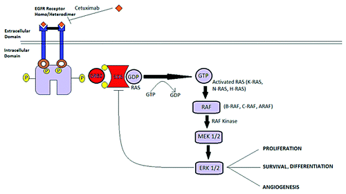
Figure 1. Pathology showed a moderately differentiated adenocarcinoma in the rectum with direct extension to the ovary and uterus at the time of recurrence (H&E, 200× original magnification).
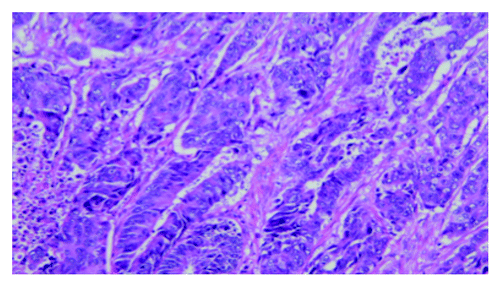
Figure 2. Tumor markers CA19-9 and CEA
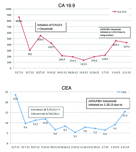
Figure 3. (A) Coronal PET image shows multiple fluoro-18-deoxyglucose (FDG) avid nodules (white arrows) scattered throughout both lungs and right hila region, consistent with thoracic metastatic disease. (B) Axial CT scan obtained at the same time show multiple bilateral pulmonary nodules (black arrows) of various sizes, ranging from a few millimeters to a couple of centimeters, consistent with pulmonary metastasis form colorectal cancer. There was no appreciable change in lesion size or FDG avidity with treatment.
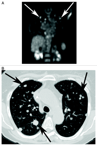
Figure 4. EGFR/KRAS pathway.