Figures & data
Figure 1. NSCLC cells express A2AR. (A) IHC analysis of A2AR expression in a lung cancer TMA. Representative pictures of 0 and 3+ A2AR expressing tumors are shown. (B) Table showing the expression of A2AR in lung tumors from the TMA. 0, no expression; +1 to +3, increasing expression of A2AR. (C) Immunoblot analysis of 8 NSCLC cell lines show expression of the A2AR.
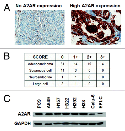
Figure 2. CAFs express A2AR. (A) IHC analysis of A2AR expression in a lung cancer TMA. Representative pictures of 0 and 2+ A2AR expressing fibroblasts are shown. Arrow shows the fibroblast in the picture. (B) Table showing the expression of A2AR in the fibroblasts of lung tumors from the TMA. 0, no expression; +1 to +3, increasing expression of A2AR. (C) Immunoblot analysis of A2AR and α-SMA in a panel of 5 CAF. Expression of (D) FAP-α and (E) CD73 were detected by flow cytometric analysis on lymphocytes (dotted line, negative control) and a panel of 5 CAF (all other lines).
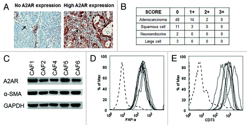
Figure 3. A2AR antagonists decrease tumor growth in a mouse xenograft model. (A) Nude mice (4‒6 wks old) were inoculated s.c. with 7.5 × 10⁶ PC9 cells in the right flank. After 1 week the tumors were palpable and treatment with vehicle control (● 15% DMSO, 15% Cremophore EL, 70% H2O), SCH58261 2 mg/kg (▲), and ZM241385 10 mg/kg (■) started. Drugs were given via i.p. injections for 20 d. (B) A significant decrease in tumor burden was observed with both ZM241385 and SCH58261 treatment.
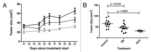
Figure 4. A2AR antagonists induce apoptotic cell death. (A) Morphological analysis PC9 cells untreated, vehicle control (DMSO), and treated with ZM241385 (25 μM; 48 h). Notice the marked decrease in adhering cells in ZM241385 treated cells. (B) A549 and PC9 cells were treated with vehicle control (DMSO) and ZM241385 (25 μM; 48 h) and the percentage of apoptotic and dead cells determined as described in Materials and Methods. ZM241385 causes significant apoptosis and cell death as compared with vehicle control (P < 0.05). Means ± SD from 6 experiments are presented. (C) Representative of an Annexin V/PI histogram. (D) PC9 cells were treated with vehicle control, ZM241385 (25 μM; 48 h), the pan-caspase inhibitor Z-VAD.fmk (50 μM; 1 h pre-treatment) and ZM241385 in the presence of Z-VAD.fmk and immunoblotting analysis of PARP cleavage was performed. ZM241385 treatment causes significant PARP cleavage, while pre-treatment with Z-VAD.fmk prevented cleavage of PARP.
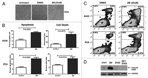
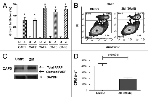
Figure 5. A2AR antagonists induce inhibition of cell proliferation. (A) CAFs were treated with vehicle control (DMSO; D) or ZM241385 (25 μM; Z). After 72 h an MTS assay was performed. ZM241385 significantly inhibited the growth in all 5 CAFs (*P < 0.05). Means +/− SEM from 3 experiments are presented. (B) CAF5 cells were treated with vehicle control (DMSO) and ZM241385 (25 μM; 96 h). ZM241385 does not cause apoptosis as compared with vehicle control as shown in the representative histogram. (C) CAF5 cells were treated with vehicle control (DMSO) and ZM241385 (25 μM; 4 h) and immunoblotting analysis of PARP cleavage was performed. ZM241385 treatment did not cause PARP cleavage. (D) Decrease in cell proliferation (3HdT assay) on CAF5 in the presence of ZM241385 (25 μM; 48 h) is significant when compared with vehicle control (DMSO). Means ± SEM from 3 experiments are presented.