Figures & data
Figure 1. (A) Brightfield photomicrograph of representative terminal lobular ductal unit (TDLUs) in unstained breast tissue section from which infrared (IR) spectra were acquired from epithelial cells (E), intralobular stroma (LS), and interlobular stroma (ILS). Tissue sections used were selected from subjects at either end of the spectrum of immunostaining for 4-hydroxy2-nonenal (4HNE adducts) adducts.Citation43 Specifically, sections in the 4HNE+ category were from subjects whose breast tissue sections showed consistently a preponderance of TDLUs occupied by many 4HNE+ ME cells, while those in the 4HNE− category were from subjects whose tissue sections showed consistently at most a few TDLUs occupied by a small number of 4HNE+ ME cells.Citation43 4HNE− and 4HNE+ TDLUs were interrogated by synchrotron radiation-based Fourier-transform IR (SR-FTIR) microspectroscopy via a 10 μm × 10 μm beam aperture either to acquire point IR spectra or in a 10 μm step-wise fashion to derive image maps The red crosses identify locations from which point spectra were acquired one after another. The software used enables tracking along the brightfield image of the tissue section and pinpoint the exact locations from which spectra were derive spectra, i.e., along the epithelial cells. Once the points of interest are identified, the spectral acquisition program is activated, a motorized stage in chronological order moves each point of interest to the focus of the synchrotron IR beam to allow for spectral acquisition. (B) Average of all the IR spectra acquired from point IR spectra from 4HNE+ and 4HNE− TDLUs associated with E, LS, or ILS showing a typical wavenumber-absorbance intensity profile.
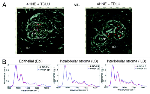
Figure 2. Principal component analysis-linear discriminant analysis (PCA-LDA) of infrared (IR) spectra of three anatomical compartments of 4-hydroxy-2-nonenal (4HNE) immunopositive (4HNE+) and 4HNE immunonegative (4HNE−) breast terminal ductal lobular units (TDLUs). Tissue sections in the 4HNE+ category were from subjects whose tissue sections showed consistently a preponderance of TDLUs occupied by many 4HNE+ ME cells, while those in the 4HNE− category were from subjects whose breast tissue sections showed consistently at most a few TDLUs occupied by a small number of 4HNE+ ME cells.Citation43 (A) PCA-LDA scores plot of point-map IR spectra (n = 540 total) of epithelial cells (EPI). Each spectral point is derived from the average of 10 IR spectra. A total of four 4HNE+ TDLUs were interrogated from two 4HNE+ sections, whereas five 4HNE− TDLUs were interrogated from three tissue sections. Discriminating wavenumbers were determined from consequent cluster vectors plots. (B) PCA-LDA scores plot of point-map IR spectra of intralobular stroma (LS; n = 90). Each spectral point is derived from the average of two IR spectra. 4HNE+ and 4HNE− TDLUs were compared as independent categories. Discriminating wavenumbers were determined from consequent cluster vectors plots. (C) PCA-LDA scores plot of point-map IR spectra of interlobular stroma (ILS; n = 180). Each spectral point is derived from the average of two IR spectra. Designated 4HNE+ and 4HNE− TDLUs were compared as independent categories. Discriminating wavenumbers were determined from consequent cluster vectors plots. Significance of category segregation was determined using an unpaired t test. Three wavenumbers contributed most to segregation of 4HNE+ vs. 4HNE− categories for a particular tissue component are listed (see each corresponding table per column).
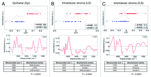
Figure 3. Unsupervised exploratory analyses by principal component analysis (PCA) of infrared (IR) spectra (1450 cm−1–900 cm−1) derived from 4-hydroxy-2-nonenal (4HNE) immunopositive (4HNE+) (n = 4) vs. 4HNE− immunonegative (4HNE−) (n = 5) terminal lobular ductal units (TDLUs). Tissue sections were categorized as 4HNE+ and 4HNE− as described in Methods and in legends to and . IR spectra were obtained from 4HNE+ and 4HNE− ME cells within the two classes of TDLUs (n = 540 total), giving n = 240 for 4HNE+ and n = 300 for 4HNE−. Spectra from 4HNE− TDLUs formed a single cluster of points in the PCA scores plot (red symbols), whereas those derived from 4HNE+ TDLUs formed two distinct clusters (blue symbols).

Figure 4. Segregating wavenumbers in the DNA/RNA region (1450 cm−1–900 cm−1) of the infrared (IR) spectrum discriminating 4-hydroxy-2-nonenal (4HNE) immunopositive (4HNE+) (n = 4) vs. 4HNE immunonegative (4HNE−) (n = 5) mammary epithelial (ME) cells in breast terminal ductal lobular units (TDLUs). Tissue sections were categorized as 4HNE+ and 4HNE− as described in Methods and in legends to and . The PCA-LDA scores plot showed good segregation between IR spectra (n = 540) derived from epithelial cells of 4HNE+ vs. 4HNE− TDLUs. From the consequent cluster vectors plot, the five wavenumbers contributing most to segregation were prioritized. To identify the most segregating wavenumbers, these isolated spectral regions were then inputted into a PCA-LDA scores plot. Significance of category segregation was determined using an unpaired t test.
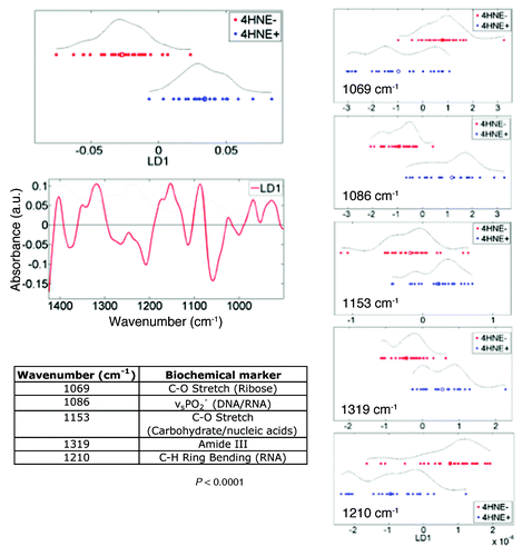
Figure 5. Unsupervised exploratory analyses by principal component analysis (PCA) of infrared (IR) spectra derived from luminal cells vs. basal, myoepithelal of cells in breast terminal ductal lobular units (TDLUs; n = 3) and extracted from image maps. A PCA scores plot shows good segregation between cells in the luminal epithelium (red symbols; n = 195) and in the basal, myoepithelium (blue symbols; n = 90) clusters. Loadings plots along principal components (PCs) were then derived in order to identify discriminating wavenumbers. Significance of category segregation along individual PCs was determined using an unpaired t-test. Distinction between 4HNE+ and 4HNE− is not needed here, as this is proof that one can separate luminal and myoepithelial cell spectra with no a priori knowledge.
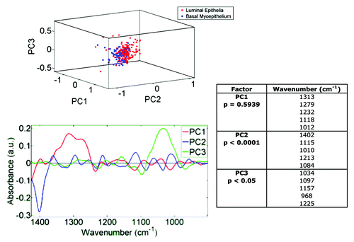
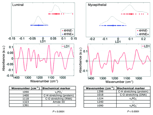
Figure 6. Principal component analysis-linear discriminant analysis (PCA-LDA) of luminal and basal, myoepithelial mammary epithelial cells in 4-hydroxy-2-nonenal (4HNE) immunopositive (4HNE+) and 4HNE immunonegative (4HNE−) breast terminal ductal lobular units (TDLUs). Tissue sections were categorized as 4HNE+ and 4HNE− as described in Methods and in legends to and . 4HNE+ and 4HNE− TDLUs (n = 3) were interrogated by synchrotron radiation-based Fourier-transform IR (SR‑FTIR) microspectroscopy via a 10 μm × 10 μm beam aperture in a 10 μm step-wise fashion to derive image maps; extracted infrared (IR) spectra of either luminal (n = 195) or basal epithelial cells (n = 90) of 4HNE+ and 4HNE− TDLUs were then obtained. Wavenumbers in the DNA/RNA region (1450 cm−1–900 cm−1) of the IR spectrum were incorporated into PCA-LDA. The PCA-LDA scores plots show good segregation between IR spectra derived from both luminal and basal epithelial cells of 4HNE+ vs. 4HNE− TDLUs. From the consequent cluster vectors plots, the five wavenumbers contributing most to segregation were prioritized. Significance of category segregation was determined using an unpaired t test.