1,725
Views
26
CrossRef citations to date
0
Altmetric
Research Paper

Maritoclax induces apoptosis in acute myeloid leukemia cells with elevated Mcl-1 expression

, , , , , , & show all
Pages 1077-1086 | Received 15 Apr 2014, Accepted 09 May 2014, Published online: 19 May 2014

Figures & data

Figure 1. Maritoclax induces Mcl-1 proteasomal degradation but not transcriptional repression. (A) U937 cells were treated with DMSO or 2.5 μM maritoclax with the indicated concentrations of MG132 for 12 h, and protein expression was analyzed by immunoblotting. (B) U937 cells were treated with DMSO or 2.5 μM maritoclax for 9 h before adding 10 µM MG132 for 3 h, and protein expression was analyzed by immunoblotting. (C) U937 cells were treated with 2.5 µM maritoclax for the indicated times, and MCL1 mRNA expression was analyzed by qRT-PCR.
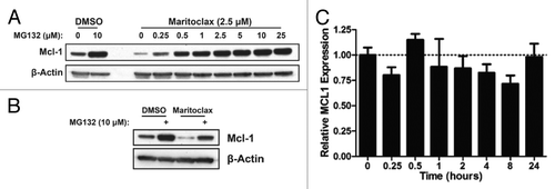
Figure 1. Maritoclax induces Mcl-1 proteasomal degradation but not transcriptional repression. (A) U937 cells were treated with DMSO or 2.5 μM maritoclax with the indicated concentrations of MG132 for 12 h, and protein expression was analyzed by immunoblotting. (B) U937 cells were treated with DMSO or 2.5 μM maritoclax for 9 h before adding 10 µM MG132 for 3 h, and protein expression was analyzed by immunoblotting. (C) U937 cells were treated with 2.5 µM maritoclax for the indicated times, and MCL1 mRNA expression was analyzed by qRT-PCR.

Figure 2. Maritoclax potency correlates with Mcl-1 expression in primary human AML. (A) The EC50 of maritoclax in 4 primary human AML samples were assayed by treating samples with maritoclax over 48 h. Error bars = SD (n = 3). (B) The expression of Bcl-2 family proteins were detected for the same 4 primary human AML samples through immunoblotting, with the Raji Burkitt lymphoma cell line as positive control. (C) Primary human AML case #555 was treated with the indicated concentrations of maritoclax for 24 h, and protein expression was analyzed by immunoblotting.
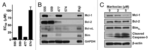
Figure 2. Maritoclax potency correlates with Mcl-1 expression in primary human AML. (A) The EC50 of maritoclax in 4 primary human AML samples were assayed by treating samples with maritoclax over 48 h. Error bars = SD (n = 3). (B) The expression of Bcl-2 family proteins were detected for the same 4 primary human AML samples through immunoblotting, with the Raji Burkitt lymphoma cell line as positive control. (C) Primary human AML case #555 was treated with the indicated concentrations of maritoclax for 24 h, and protein expression was analyzed by immunoblotting.

Figure 3. Maritoclax induces apoptosis through Mcl-1 degradation in Mcl-1-dependent AML cell lines. (A) The Bcl-2 family protein expression for a number of parental and drug-resistant AML cell lines. (B) The effective concentration for 50% viability (EC50) of parental and drug-resistant AML cell lines in response to ABT-737 and maritoclax treatment. (C) Detection of Mcl-1 degradation and caspase activation by immunoblotting in the HL60/ABTR cell line with 2 µM maritoclax over the indicated time. (D) HL60/ABTR (top) and KG1a/ABTR (bottom) cell lines were treated with a single concentration of maritoclax (2 and 1 µM, respectively) and the indicated concentrations of ABT-737 to measure viability. Error bars = SD (n = 3).
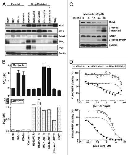
Figure 3. Maritoclax induces apoptosis through Mcl-1 degradation in Mcl-1-dependent AML cell lines. (A) The Bcl-2 family protein expression for a number of parental and drug-resistant AML cell lines. (B) The effective concentration for 50% viability (EC50) of parental and drug-resistant AML cell lines in response to ABT-737 and maritoclax treatment. (C) Detection of Mcl-1 degradation and caspase activation by immunoblotting in the HL60/ABTR cell line with 2 µM maritoclax over the indicated time. (D) HL60/ABTR (top) and KG1a/ABTR (bottom) cell lines were treated with a single concentration of maritoclax (2 and 1 µM, respectively) and the indicated concentrations of ABT-737 to measure viability. Error bars = SD (n = 3).

Figure 4. Maritoclax induces leukemic cell death in co-culture with HS-5 stroma while sparing stroma cells. U937-luc cells were cultured with or without HS-5 stroma cells and treated with maritoclax (A) or daunorubicin (B) for 48 h, and luciferase activity was measured. (C) HS-5 cells were treated with daunorubicin or maritoclax for 48 h, and viability was assayed. Error bars = SD (n = 3).
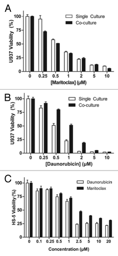
Figure 4. Maritoclax induces leukemic cell death in co-culture with HS-5 stroma while sparing stroma cells. U937-luc cells were cultured with or without HS-5 stroma cells and treated with maritoclax (A) or daunorubicin (B) for 48 h, and luciferase activity was measured. (C) HS-5 cells were treated with daunorubicin or maritoclax for 48 h, and viability was assayed. Error bars = SD (n = 3).

Figure 5. Maritoclax spares bone marrow while inducing apoptosis in mouse AML C1498 through Mcl-1 degradation. (A) Mouse AML cell line C1498 was treated with the indicated concentrations of maritoclax, ABT-737, or daunorubicin to measure cell viability. Error bars = SD (n = 3). (B) C1498 cells were treated with the indicated concentrations of maritoclax or ABT-737 for 24 h, and protein expression was analyzed by immunoblotting. (C) An in vitro culture of primary mouse bone marrow was treated with the indicated concentrations of maritoclax, ABT-737, or daunorubicin for 48 h to measure cell viability. Error bars = SD (n = 3). (D) Primary mouse bone marrow was seeded in methylcellulose medium to allow for progenitor cell growth, and treated with the indicated concentrations of maritoclax or daunorubicin for 7 d, and the CFU-GEMM, CFU-GM, BFU-E, and CFU-E colonies were counted.
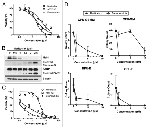
Figure 5. Maritoclax spares bone marrow while inducing apoptosis in mouse AML C1498 through Mcl-1 degradation. (A) Mouse AML cell line C1498 was treated with the indicated concentrations of maritoclax, ABT-737, or daunorubicin to measure cell viability. Error bars = SD (n = 3). (B) C1498 cells were treated with the indicated concentrations of maritoclax or ABT-737 for 24 h, and protein expression was analyzed by immunoblotting. (C) An in vitro culture of primary mouse bone marrow was treated with the indicated concentrations of maritoclax, ABT-737, or daunorubicin for 48 h to measure cell viability. Error bars = SD (n = 3). (D) Primary mouse bone marrow was seeded in methylcellulose medium to allow for progenitor cell growth, and treated with the indicated concentrations of maritoclax or daunorubicin for 7 d, and the CFU-GEMM, CFU-GM, BFU-E, and CFU-E colonies were counted.

Figure 6. Maritoclax is effective in vivo against U937 xenografts in athymic nude mice. (A) The tumor volumes of nude mice bearing U937 xenograft through daily treatment with vehicle or 20 mg/kg maritoclax. Error bars = SD. (B) The body weight of the same nude mice throughout the course of treatment, expressed as percent change relative to initial treatment. (C) Histological analyses of the heart sections by H&E and Masson trichrome from a representative nude mouse from each treatment arm 16 d after initial treatment.
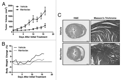
Figure 6. Maritoclax is effective in vivo against U937 xenografts in athymic nude mice. (A) The tumor volumes of nude mice bearing U937 xenograft through daily treatment with vehicle or 20 mg/kg maritoclax. Error bars = SD. (B) The body weight of the same nude mice throughout the course of treatment, expressed as percent change relative to initial treatment. (C) Histological analyses of the heart sections by H&E and Masson trichrome from a representative nude mouse from each treatment arm 16 d after initial treatment.

Table 1. Classification of tumor response to maritoclax 17 d following treatment termination

Table 2. The complete blood count (mean ± SEM) from mice treated with vehicle or maritoclax for 16 d

Supplemental material

Additional material

Download Zip (287.5 KB)

Reprints and Corporate Permissions

Please note: Selecting permissions does not provide access to the full text of the article, please see our help page How do I view content?

To request a reprint or corporate permissions for this article, please click on the relevant link below:

Academic Permissions

Please note: Selecting permissions does not provide access to the full text of the article, please see our help page How do I view content?

Obtain permissions instantly via Rightslink by clicking on the button below:

If you are unable to obtain permissions via Rightslink, please complete and submit this Permissions form. For more information, please visit our Permissions help page.