Figures & data
Figure 1. Treatment with combination therapy results in the lowest proliferation rates and highest levels of apoptosis. (A) MCF7/V and (B) MCF7/CD1 cells were treated over the course of 9 d with control DMSO, 400 nM CDK4i, 30 nM doxorubicin, or combination therapy, and an MTS assay was used to measure cell number at the indicated time points. Absorbance for CDK4i, doxorubicin, and combination treated cells was compared with control treated cells within each cell line using a Student t test to obtain P values. Absorbance values are shown ± SE. (C) MCF7/V and (D) MCF7/CD1 cells were treated over the course of 2 d with control DMSO, 400 nM CDK4i, 30 nM doxorubicin, or combination therapy. Cells were fixed and stained using a TUNEL assay and cells undergoing apoptosis were counted and normalized to control treated cells to obtain fold changes. Experiments were repeated 3 times and representative results are shown. * indicates P < 0.05.
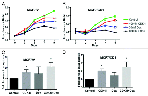
Figure 2. Treatment with CDK4i and doxorubicin combination therapy or transduction with 5M Smad3 suppresses colony growth. (A) MCF7/V and MCF7/CD1 cells were transduced with CS2 vector, WT Smad3, or 5M Smad3. Protein was extracted and immunoblotted with Smad3, cyclin D, and GAPDH. (B) MCF7/V and (C) MCF7/CD1 cells were transduced with CS2 vector control, WT Smad3, or 5M Smad3 and treated over the course of 12 d with control DMSO, 400 nM CDK4i, 30 nM doxorubicin, or combination therapy. Colony area was measured at the indicated time points using Metamorph software. Experiments were repeated 3 times and representative results are shown. Error bars indicate ± SE. * indicates P < 0.05 for comparisons between vector transduced cells and WT and 5M transduced cells treated with control. ^ indicates P < 0.05 for comparisons between control treatment and study treatments for each transduced construct within each cell line.
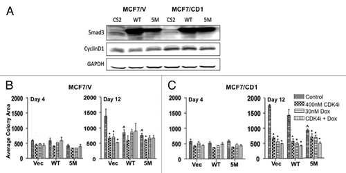
Figure 3. The array shows that CDK4i and doxorubicin therapy affect expression of proteins associated with apoptosis. (A) MCF7/V and (B) MCF7/CD1 were treated with control DMSO, 400 nM CDK4i, 30 nM doxorubicin, or combination therapy. Protein was collected and applied to the array. Protein expression was quantified using Multigauge software. The array was repeated 3 times and representative results are shown. Error bars indicate ± SE.
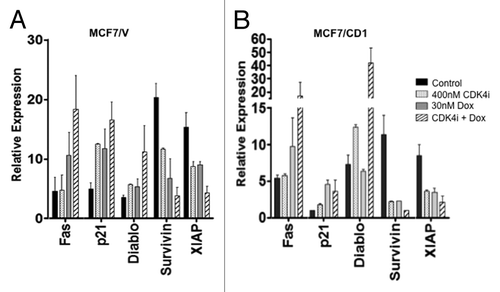
Figure 4. Survivin expression decreases upon treatment with CDK4 inhibitor and doxorubicin alone or in combination. (A) MCF7/V and MCF7/CD1 cells were treated with DMSO, 400 nM CDK4i, 30 nM doxorubicin, or combination treatment for 48 h. Protein was extracted, followed by immunoblotting with survivin, XIAP, Smac/DIABLO, p21, pRb, and β-Actin antibodies. (C) MCF7/V and MCF7/CD1 cells were treated with DMSO, 400 nM CDK4i, 30 nM doxorubicin, or combination treatment for 48 h. Mitochondrial and cytoplasmic fractions were isolated, followed by immunoblotting with Smac/DIABLO, Cox IV, and GAPDH antibodies. Experiments were repeated 3 times and representative results are shown. * indicates P < 0.05.
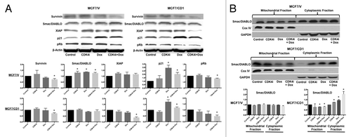
Figure 5. Intact Smad3 signaling and doxorubicin treatment result in the greatest reduction in survivin expression. (A) MCF7/V and (B) MCF7CD1 cells were transduced with CS2 vector control, WT Smad3, or 5M Smad3 and then treated with either DMSO control or 30 nM doxorubicin for 48 h. Protein was extracted, followed by immunoblotting with survivin, XIAP, Smac/DIABLO, and β-actin antibodies. Experiments were repeated 3 times and representative results are shown. * indicates P < 0.05.
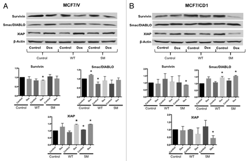
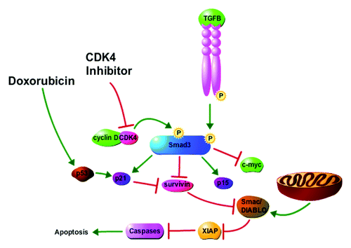
Figure 6. Proposed model for impact of CDK4i and doxorubicin on cancer cell cycle arrest and apoptosis. Cyclin D overexpression mediates CDK4 phosphorylation of Smad3, resulting in lower expression of the Smad3-regulated cdkis p15 and p21 and higher expression of c-myc and survivin. Increased survivin levels inhibit apoptosis through the regulation of Smac/DIABLO and XIAP. CDK4i treatment results in cell cycle arrest and induction of apoptosis through inhibition of CDK4-mediated phosphorylation of Smad3 and the consequent increase of p15 and p21 expression and decrease of c-myc and survivin expression. Doxorubicin treatment results in cell cycle arrest and apoptosis, in part, through the induction of p21 and the direct action of p21 on inhibition of survivin. The figure was made with tools from http://www.proteinlounge.com.