Figures & data
Figure 1. Mechanistically distinct chemotherapeutic drugs caused the release of mature IL-1β by BMDM. After serum deprivation for 0.5 h, BMDM were pre-treated with 50 ng/mL LPS for 4 h and then washed away. The cells were then treated with the indicated chemotherapeutic drugs for 18 h. LPS-primed BMDM were treated with medium alone (lane 1), azacitidine (lane 2), cisplatin (lane 3), cytarabine (lane 4), etoposide (lane 5), fludarabine (lane 6), melphalan (lane 7), methotrexate (lane 8), paclitaxel (lane 9), vincristine (lane 10), 5-FU (lane 11), or doxorubicin (lane 12). Western blots of cell lysates and medium samples were then processed using antibodies against total p38 MAPK in the cell lysates (as loading control) or IL-1β in the medium.
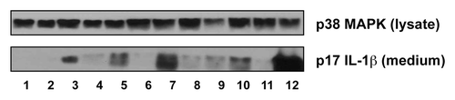
Figure 2. Doxorubicin and vincristine synergistically induce IL-1β production. BMDM from WT or mutant mice were pre-treated LPS for 4 h and then with doxorubicin, vincristine, or both for 18 h. Western blots of cell lysates and medium samples were then processed using antibodies against total p38 MAPK in the cell lysates (as loading control) or IL-1β in the medium. (A) LPS-primed or unprimed BMDM were treated with doxorubicin, vincristine, or both as indicated. (B) LPS-primed or unprimed BMDM from WT or Nlrp3−/− mice were treated with doxorubicin and vincristine as indicated. (C) LPS-primed BMDM from WT or Tnfr1−/− Il1r1−/− mice were treated with doxorubicin, vincristine, or both as indicated.
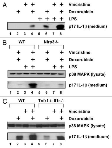
Figure 3. Doxorubicin and vincristine synergistically increased gene expression. RNA was extracted from LPS-unprimed BMDM after 12 h of exposure to doxorubicin, vincristine, or both in the presence or absence of anakinra. The expression of IL-1β, IL-6, and CXCL1 was quantitated using real-time RT-PCR.
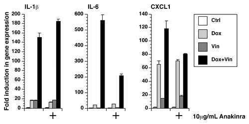
Figure 4. Doxorubicin and vincristine affects incorporation of leucine. Cells were exposed to doxorubicin, vincristine, or both for various times as indicated. [3H]-leucine was added for the final 30 min. The level of radioactive incorporation was quantitated using liquid scintillation.
![Figure 4. Doxorubicin and vincristine affects incorporation of leucine. Cells were exposed to doxorubicin, vincristine, or both for various times as indicated. [3H]-leucine was added for the final 30 min. The level of radioactive incorporation was quantitated using liquid scintillation.](/cms/asset/b0f68fff-df48-4256-bee6-121ad9c51668/kcbt_a_10929922_f0004.gif)
Figure 5. Small-molecule kinase inhibitors block the expression of IL-1β, IL-6, and CXCL1. BMDM were treated with doxorubicin, vincristine, or both for 12 h in the presence or absence of nilotinib, ponatinib, or sorafenib as indicated. (A) RNA was extracted from the cells and processed for real-time RT-PCR. (B) Cytokine levels in the medium were quantitated using a multiplex assay.
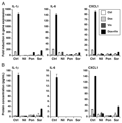
Figure 6. Doxorubicin and vincristine activated p38 MAPK through ZAK. BMDM from WT or Zak−/− mice were treated with doxorubicin, vincristine, or both for 12 h. Western blots of cell lysates were processed using antibodies against phosphorylated JNK, phosphorylated p38 MAPK, or total p38 MAPK.
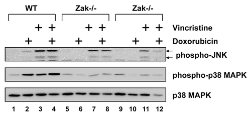
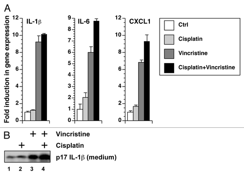
Figure 7. Cisplatin and vincristine fail to show a synergistic effect in gene expression or IL-1β processing. (A) RNA was extracted from BMDM exposed to cisplatin, vincristine, or both for 12 h. Gene expression was quantitated using real-time RT-PCR. (B) LPS-primed BMDM were treated with cisplatin, vincristine, or both. Levels of IL-1β in the medium were detected using western blotting.