Figures & data
Figure 1. The effect of zinc treatment on the cellular accumulation of zinc and proliferation of Panc1 cells. The cells were treated with zinc for 24 h. (A) Pyrithione (1 uM) was included in the medium. (B) Clioquinol (10 uM) was included in the medium.
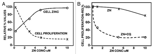
Figure 2. ZIP3 transporter and zinc treatment effects on cell proliferation. (A) Immunohistochemical identification of ZIP3 transporter in adenocarcinoma and normal human pancreatic tissue sections. Blue arrows show basal membrane localization of transporter in normal ductal and acinar epithelium. Black arrows show the absence of plasma membrane transporter in ductal adenocarcinoma and in PanIN epithelium. (B) Blue arrows show the presence of plasma membrane ZIP3 transporter in Panc1 cells. (C) Shows the effect of zinc treatment on proliferation of wildtype Panc1 cells that exhibit ZIP3 transporter and on Panc1 cells with downregulated ZIP3 (i.e., Panc1/ZIP3 cells vs. Panc1/-ZIP3 cells).
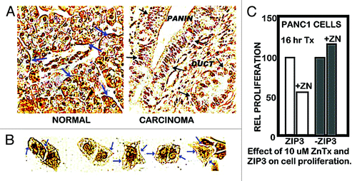
Figure 3. RREB1 in normal vs. adenocarcinoma pancreas and its regulation of ZIP3 and zinc effects on proliferation of Panc1 cells. (A) Immunohistochemistry showing abundant RREB1 in normal ductal and acinar epithelium; and marked decrease of RREB1 (especially nuclear RREB1) in well differentiated malignancy and in the PanIN epithelium. (B) Western blot of effects of overexpression of RREB1 on ZIP3 expression. Lanes are duplicates for pcDNA and for RREB1. (C) The effects of RREB1 expression of the Panc1 cells on cell proliferation in the presence of zinc.
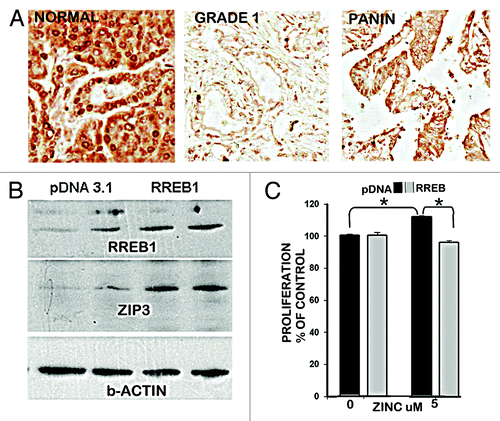
Figure 4. In situ staining of zinc levels in human pancreatic normal and adenocarcinoma tissues. (A) Low power tissue arrays showing marked decreased dithizone zinc staining in adenocarcinoma. (B) Enlargement to show the high zinc in the normal ductal and acini epithelium vs. zinc depletion in adenocarcinoma. (C) Shows the marked loss of zinc in PanIN and in the surrounding ductal adenocarcinoma. (D) Zinquin staining showing decreased zinc (loss of fluorescence) in adenocarcinoma.
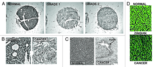
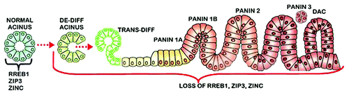
Figure 5. The concept of the “RREB1/ZIP3/Decreased Zinc” transformation and acini de-differentiation as the origin of PanlN leading to the development of ductal adenocarcinoma. De-diff, de-differentiation; Trans-diff, trans-differentiation; DAC, ductal adenocarcinoma. (The figure is a modified version from Morris et al.Citation31)