Figures & data
Figure 1 DNA damage reduces MDM2 reactivity to SMP14 antibody. (A) Diagram of MDM2 and epitope locations of the monoclonal antibodies used in this study. (B) U2OS osteosarcoma cells were treated with 10 Gy IR and analyzed for MDM2 level at indicated time points by western blot using 3G9. Identical amount of whole cell extract was used in each lane. (C and D) U2OS and normal human fibroblasts were treated with 10 Gy IR and analyzed for MDM2 level at indicated time points by western blot using different antibodies. (E) U2OS cells were incubated with Neocarzinostatin and analyzed for MDM2 level at indicated time points by western blot.
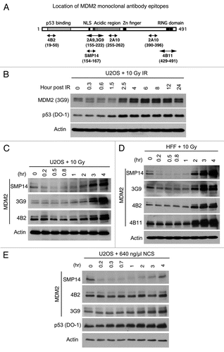
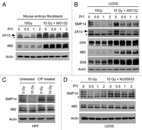
Figure 2 Loss of SMP14 signal after DNA damage due to epitope masking. (A and B) Mouse embryo fibroblasts and U2oS were treated with 10 Gy IR in the absence or presence of 30 µM MG132. MDM2 level was analyzed by western blot at indicated time points using 2A10. Arrow indicates MDM2 band detected by 2A10. A background band is marked by *. (C) Normal human fibroblasts were irradiated with 10 Gy and after 1 h whole cell extract was treated with phosphatase (CIP) or incubated without CIP. Samples were analyzed by SMP14 western blot. The membrane was stripped and reprobed using 4B2. (D) U2OS cells were irradiated with 10 Gy in the absence or presence of 10 µM ATM inhibitor KU55933. Samples were analyzed by SMP14 western blot. A background band is marked by *.