Figures & data
Figure 1 Scatterplots of normalized gene expression levels for each transcript in individual oocytes mature at retrieval (group 1) or maturing post-retrieval (group 2). Bars represent the median level.
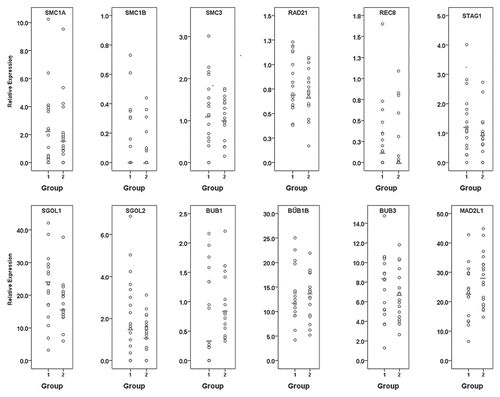
Figure 2 Incidence of relative gene expression levels in individual rhesus macaque oocytes mature at retrieval (group 1; dark gray bars) or maturing post-retrieval (group 2; light gray bars). (A) Quantity of oocytes lacking each specific transcript; (B) Quantity of oocytes with specific transcript expression levels below the median; (C) Quantity of oocytes with specific transcript expression levels above the median.*represents a statistically significant difference between group 1 and group 2 (α = 0.05).
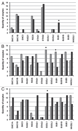
Figure 3 Correlation curves for BUB1 compared with minimal expression of shugoshins or mitotic cohesins (STAG1, SMC1A, SMC3, RAD21, SGOL1 and SGOL2). (A) significant positive correlation (rho = 0.60, p < 0.01) within group 1 oocytes. (B) Absence of a significant correlation (rho = -0.20, p = 0.43) in group 2 oocytes.
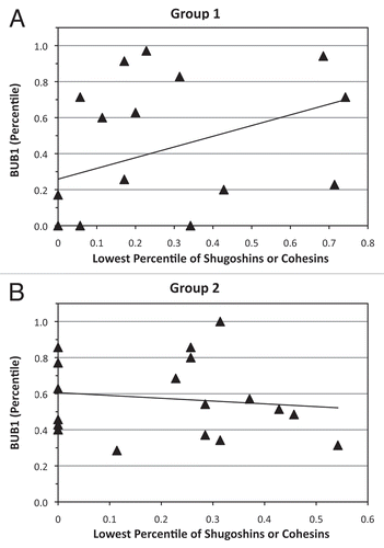
Table 1 Selected proteins associated with chromosome segregation
Table 2 Significant Correlation Coefficients between transcript expression levels in group 1 and group 2 oocytes
Table 3 Sequences of real-time PCR primers, TaqMan probe and product size