1,182
Views
20
CrossRef citations to date
0
Altmetric
Extra Views

Selective blockade of tumor angiogenesis

&
Pages 2253-2259 | Published online: 15 Jun 2012

Figures & data

Figure 1. TEM8 structure and antibody binding. (A) TEM8 is a type I membrane protein containing a vWF type A (VWA) domain in its extracellular domain. (B) The image depicts the selective binding of anti-TEM8 blocking antibodies (blue) to TEM8 receptors (orange) that are overexpressed on the surface of tumor endothelium. Tumor cells are depicted in yellow.
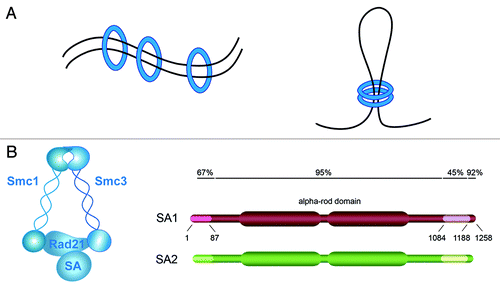
Figure 1. TEM8 structure and antibody binding. (A) TEM8 is a type I membrane protein containing a vWF type A (VWA) domain in its extracellular domain. (B) The image depicts the selective binding of anti-TEM8 blocking antibodies (blue) to TEM8 receptors (orange) that are overexpressed on the surface of tumor endothelium. Tumor cells are depicted in yellow.

Figure 2. TEM8 antibodies recognize proteins in both Tem8 WT and KO mice. (A) The rabbit anti-TEM8 polyclonal antibody from AbCAM (cat # ab19387) generated against the TEM8 extracellular domain peptide LMKLTEDREQIRQGLE was used to detect TEM8 in various normal tissues from Tem8 WT and KO mice. Although the polyclonal antibody recognized both mouse TEM8 (mTEM8) and human TEM8 (hTEM8) in 293 cells transfected with the corresponding Tem8 genes (asterisk), when tested against lysates from various normal mouse tissues it reacted predominantly with proteins that were present in both the Tem8 WT and KO mice. (B) PCR genotyping assay used to confirm the Tem8 WT and KO status of the samples used in A.
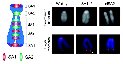
Figure 2. TEM8 antibodies recognize proteins in both Tem8 WT and KO mice. (A) The rabbit anti-TEM8 polyclonal antibody from AbCAM (cat # ab19387) generated against the TEM8 extracellular domain peptide LMKLTEDREQIRQGLE was used to detect TEM8 in various normal tissues from Tem8 WT and KO mice. Although the polyclonal antibody recognized both mouse TEM8 (mTEM8) and human TEM8 (hTEM8) in 293 cells transfected with the corresponding Tem8 genes (asterisk), when tested against lysates from various normal mouse tissues it reacted predominantly with proteins that were present in both the Tem8 WT and KO mice. (B) PCR genotyping assay used to confirm the Tem8 WT and KO status of the samples used in A.

Figure 3. Amino acid starvation induces TEM8 expression. (A) Real-time quantitative PCR revealed a gradual increase in TEM8 mRNA expression following amino acid (AA) depletion from the growth medium. (B) Immunoblotting (IB) with anti-TEM8 antibodies (clone SB5) revealed an increase in TEM8 protein 24 h following removal of amino acids from the growth medium. β actin was used as a loading control.
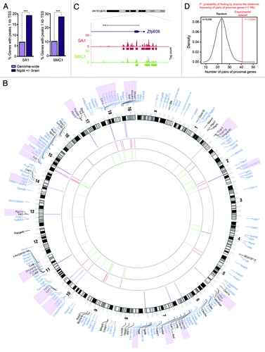
Figure 3. Amino acid starvation induces TEM8 expression. (A) Real-time quantitative PCR revealed a gradual increase in TEM8 mRNA expression following amino acid (AA) depletion from the growth medium. (B) Immunoblotting (IB) with anti-TEM8 antibodies (clone SB5) revealed an increase in TEM8 protein 24 h following removal of amino acids from the growth medium. β actin was used as a loading control.

Reprints and Corporate Permissions

Please note: Selecting permissions does not provide access to the full text of the article, please see our help page How do I view content?

To request a reprint or corporate permissions for this article, please click on the relevant link below:

Academic Permissions

Please note: Selecting permissions does not provide access to the full text of the article, please see our help page How do I view content?

Obtain permissions instantly via Rightslink by clicking on the button below:

If you are unable to obtain permissions via Rightslink, please complete and submit this Permissions form. For more information, please visit our Permissions help page.