Figures & data
Figure 1. Spatio-temporal dynamics of phospho-AMPKαThr172 during mitosis and cytokinesis in glucose-starved A431 epidermoid cancer cells. Figure shows representative portions of glucose-starved (18 h), cell dividing-containing images captured on a BD PathwayTM 855 Bioimager System with a 40x objective in different channels for fosfo-AMPKαThr172 (green) and Hoechst 33258 (blue) and merged using BD AttovisionTM software. A rabbit anti-phospho-AMPK (Thr172) polyclonal antibody (sc-101630, Santa Cruz Biotechnology) was used in this set of images (and also in –)
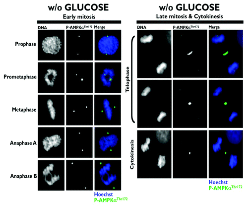
Figure 2. Co-localization analysis of phospho-AMPKαThr172 and phospho-MRLCSer19 during mitosis and cytokinesis in glucose-supplemented A431 epidermoid cancer cells. Figure shows representative portions of cell dividing-containing images captured on a BD PathwayTM 855 Bioimager System with a 40x objective in different channels for phospho-AMPKThr172 (red), phospho-MRCLSer19 (green) and Hoechst 33258 (blue) and merged using BD AttovisionTM software. The rectangular regions (white line) are enlarged and shown as high magnification insets. A phospho-myosin light chain 2 (Ser19) mouse monoclonal antibody (#3675) was used in the set of images (and also in and ).
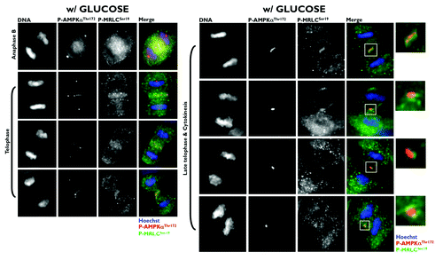
Figure 3. Co-localization analysis of phospho-AMPKαThr172 and phospho-MRLCSer19 during mitosis and cytokinesis in glucose-starved A431 epidermoid cancer cells. Figure shows representative portions of glucose-starved (18 h), cell dividing-containing images captured on a BD PathwayTM 855 Bioimager System with a 40x objective in different channels for phospho-AMPKThr172 (red), phospho-MRCLSer19 (green) and Hoechst 33258 (blue) and merged using BD AttovisionTM software. The rectangular regions (white line) are enlarged and shown as high magnification insets.
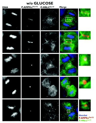
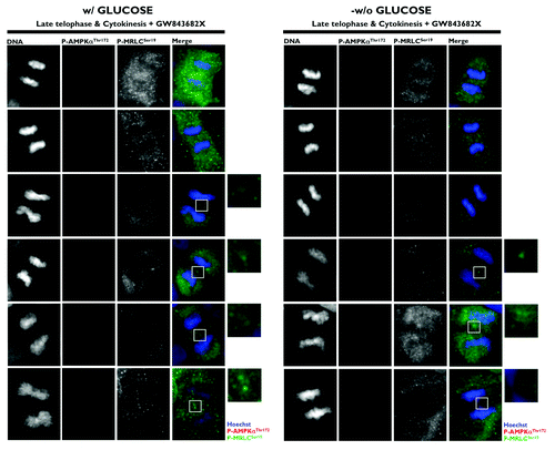
Figure 4. Spatio-temporal dynamics of phospho-AMPKαThr172 and phospho-MRLCSer19 during mitosis and cytokinesis: Impact of the PLK1 inhibitor GW843682X. Glucose-supplemented (left panels) and glucose-starved (right panels) A431 cells were treated with 10 μmol/L GW843682X for 30 min. Figure shows representative portions of cell dividing-containing images captured on a BD PathwayTM 855 Bioimager System with a 40x objective in different channels for phospho-AMPKThr172 (red), phospho-MRCLSer19 (green) and Hoechst 33258 (blue) and merged using BD AttovisionTM software. The rectangular regions (white line) are enlarged and shown as high magnification insets.