1,710
Views
50
CrossRef citations to date
0
Altmetric
Review

Phospho-specific Smad3 signaling

Impact on breast oncogenesis

&
Pages 2443-2451 | Published online: 01 Jul 2012

Figures & data

Figure 1. Canonical TGFβ signaling. TβRII phosphorylates TβRI in response to TGFβ binding. Activated TβRI phosphorylates Smad3 at its C-terminus, which releases it from the anchoring protein SARA and permits formation of a Smad3-Smad4 complex. This heteromeric complex then translocates to the nucleus, associates with DNA-binding co-activators and co-repressors and regulates the expression of target genes. I-Smads antagonize TGFβ signaling by preventing phosphorylation of Smad3 and thus act as a negative feedback mechanism. PPM1A is a phosphatase that can dephosphorylate Smad3 phosphorylated at its C-terminal SXS motif.
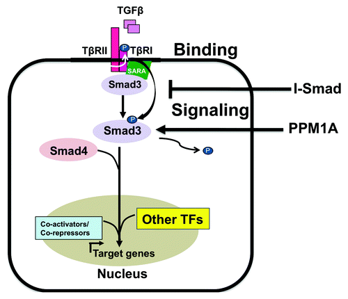
Figure 1. Canonical TGFβ signaling. TβRII phosphorylates TβRI in response to TGFβ binding. Activated TβRI phosphorylates Smad3 at its C-terminus, which releases it from the anchoring protein SARA and permits formation of a Smad3-Smad4 complex. This heteromeric complex then translocates to the nucleus, associates with DNA-binding co-activators and co-repressors and regulates the expression of target genes. I-Smads antagonize TGFβ signaling by preventing phosphorylation of Smad3 and thus act as a negative feedback mechanism. PPM1A is a phosphatase that can dephosphorylate Smad3 phosphorylated at its C-terminal SXS motif.

Figure 2. TGFβ/Smad3 signaling and the cell cycle. CyclinD/CDK4/6 and Cyclin E/CDK2 mediate the transition of the cell cycle from the G1 to S phase by phosphorylating Rb and thus preventing it from sequestering E2F. E2F is then free to drive the transcription of G1/S phase transition proteins. TGFβ/Smad3 signaling promotes cell cycle arrest through its induction of p15 and p21, in conjunction with Sp1 and repression of c-myc, in conjunction with E2F4/5 and p107. Ras, in cooperation with Pin1, can increase the transcriptional activity of cyclin D, inducing tumor promotion.
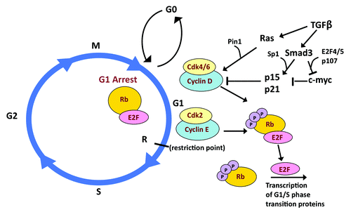
Figure 2. TGFβ/Smad3 signaling and the cell cycle. CyclinD/CDK4/6 and Cyclin E/CDK2 mediate the transition of the cell cycle from the G1 to S phase by phosphorylating Rb and thus preventing it from sequestering E2F. E2F is then free to drive the transcription of G1/S phase transition proteins. TGFβ/Smad3 signaling promotes cell cycle arrest through its induction of p15 and p21, in conjunction with Sp1 and repression of c-myc, in conjunction with E2F4/5 and p107. Ras, in cooperation with Pin1, can increase the transcriptional activity of cyclin D, inducing tumor promotion.

Figure 3. Smad3 phosphorylation sites. Smad3 is phosphorylated by TβRI at the C-terminus and by various cancer-associated kinases at indicated sites. The MH1 domain is responsible for DNA binding, while the MH2 domain mediates interactions with co-activators and co-repressors.
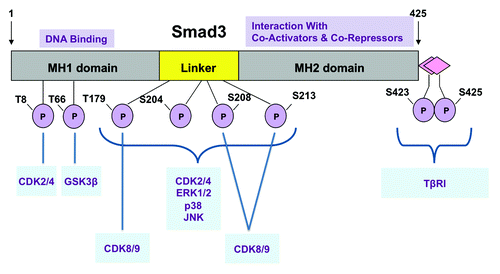
Figure 3. Smad3 phosphorylation sites. Smad3 is phosphorylated by TβRI at the C-terminus and by various cancer-associated kinases at indicated sites. The MH1 domain is responsible for DNA binding, while the MH2 domain mediates interactions with co-activators and co-repressors.

Figure 4. Model for dichotomous Smad3 signaling. Differential phosphorylation of Smad3 results in the induction of TGFβ-mediated tumor suppressive genes in early-stage breast cancer cells or oncogenic genes in late-stage breast cancer cells. Nuclear kinases CDKs 2/4 phosphorylate Smad3 after pSmad3C translocates into the nucleus, while cytoplasmic MAPKs can phosphorylate Smad3 inducing nuclear activation independent of TGβRI phosphorylation.
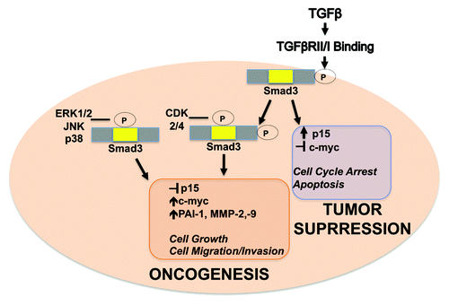
Figure 4. Model for dichotomous Smad3 signaling. Differential phosphorylation of Smad3 results in the induction of TGFβ-mediated tumor suppressive genes in early-stage breast cancer cells or oncogenic genes in late-stage breast cancer cells. Nuclear kinases CDKs 2/4 phosphorylate Smad3 after pSmad3C translocates into the nucleus, while cytoplasmic MAPKs can phosphorylate Smad3 inducing nuclear activation independent of TGβRI phosphorylation.

Table 1. Potential drug candidates that promote TGFβ-mediated tumor suppression

Reprints and Corporate Permissions

Please note: Selecting permissions does not provide access to the full text of the article, please see our help page How do I view content?

To request a reprint or corporate permissions for this article, please click on the relevant link below:

Academic Permissions

Please note: Selecting permissions does not provide access to the full text of the article, please see our help page How do I view content?

Obtain permissions instantly via Rightslink by clicking on the button below:

If you are unable to obtain permissions via Rightslink, please complete and submit this Permissions form. For more information, please visit our Permissions help page.