Figures & data
Figure 1. KLF5 promotes proliferation in cells with wild-type p53 but inhibits cell proliferation with mutant p53. (A and B) In EPC2-T cells, by MTT assay, KLF5 overexpression had little effect on cell proliferation (A), likely due to the effects of endogenous KLF5, but proliferation was significantly reduced by KLF5 suppression with shRNA (B). (C and D) In contrast, in EPC2-T-p53R175H cells, KLF5 induction inhibited cell proliferation (C), while KLF5 knockdown increased proliferation (D). EV, empty vector control; * p < 0.05, ** p < 0.005
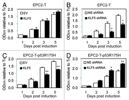
Figure 2. KLF5 upregulates the cyclin-dependent kinase inhibitor p21Waf1/Cip1 in cells with mutant p53. (A and B) When KLF5 was induced in EPC2-T cells, which contain wild-type p53, p21Waf1/Cip1 protein (A), by western blot, and mRNA (B), by quantitative real-time PCR, were unchanged, similar to the effects on proliferation seen with KLF5 induction. In contrast, KLF5 knockdown by shRNA, increased p21Waf1/Cip1 in EPC2-T cells. (C and D) In EPC2-T-p53R175H cells, p21Waf1/Cip1 was normally expressed at low levels, and KLF5 overexpression increased p21Waf1/Cip1 protein (C) and mRNA (D), while KLF5 suppression reduced p21Waf1/Cip1. EV, empty vector control; *p < 0.05, ** p < 0.01
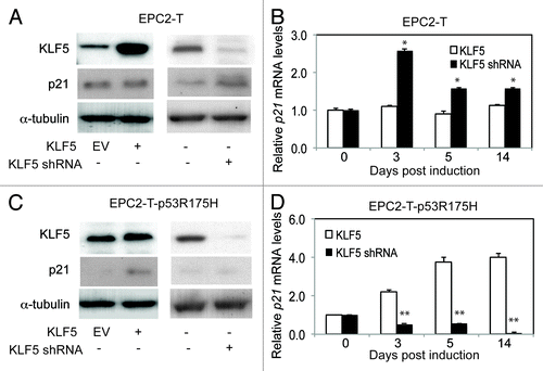
Figure 3. KLF5 directly binds and transactivates p21Waf1/Cip1 in the presence of p53R175H. (A) The 5′ upstream regulatory region of p21Waf1/Cip1 contains two putative KLF5 binding sites (underlined) within two p53 response elements (p53RE1 and p53RE2). (B) Quantitative ChIP revealed that KLF5 binding on p21Waf1/Cip1 was markedly increased in cells with p53R175H as compared with cells with wild-type p53. (C) A reciprocal pattern was seen for p53 binding to p21Waf1/Cip1. (D) Luciferase reporter assays revealed that KLF5 repressed p21Waf1/Cip1 in EPC2-T cells but was a transcriptional activator of p21Waf1/Cip1 in EPC2-T-p53R175H cells. *p < 0.05, ** p < 0.01
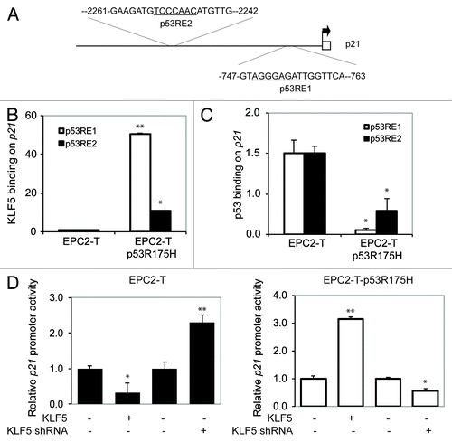
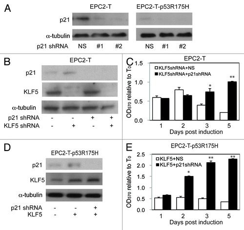
Figure 4. p21Waf1/Cip1 mediates the p53-dependent effects of KLF5 on cell proliferation. (A) By western blot, p21Waf1/Cip1 shRNA #1 and #2 successfully decreased p21Waf1/Cip1, compared with non-silencing (NS) control. (B and C) In EPC2-T cells with p21Waf1/Cip1 and/or KLF5 knockdown (B), p21Waf1/Cip1 suppression prevented the growth-inhibitory effect of KLF5 knockdown (C). (D and E) EPC2-T-p53R175H cells with KLF5 induction and/or p21Waf1/Cip1 suppression (D) demonstrated that p21Waf1/Cip1 knockdown prevented the growth-inhibitory effect of KLF5 expression in these cells. *p < 0.05, ** p < 0.01