Figures & data
Figure 1. pVHL suppresses ER-α. (A) Expression of ER-α in VHL-deficient C2 cells. MCF7 and MDA-MB.468 (MDA) cells were used as positive and negative controls of ER-α expression. Actin was used as a loading control. (B) pVHL suppresses ER-α. After transfection with the indicated vectors or si-RNAs for 24 h, 293 cells were treated with estrogen (Est; 600 μg/ml) for 6 h. The expression of ER-α was determined by an ER-α-specific Ab, and the expression of exogenous pVHL was determined by a FLAG-Ab. EV indicates an empty vector transfection. LE and SE indicate a long exposure and a short exposure, respectively. (C) The reduction of ER-α and pVHL. Two hundred and ninety-three cells were transfected with VHL and/or ER-α for 24 h. After fixation with Me-OH, 293 cells were stained with anti-ER-α (green), anti-pVHL (red) and DAPI (blue). (D) pVHL suppresses ER-α in pVHL-mutant Caki-2 cells. Caki-2 cells were transfected with the indicated vectors for 24 h and were treated with estrogen for 6 h. (E) pVHL suppresses endogenous ER-α. MCF7 cells were transfected with the indicated vectors or si-RNAs for 24 h and were treated with estrogen for 6 h. LE and SE indicate a long exposure and a short exposure, respectively. (F) The reduction of endogenous ER-α by VHL transfection. MCF-7 cells were transfected with VHL or HIF-1α for 24 h. After fixation with Me-OH, MCF7 cells were stained with anti-ER-α (green) and DAPI (blue). To check for any effects that could have been caused by the serum, cells were incubated in serum-free medium (SF) or complete medium (CM) for 6 h before harvest.
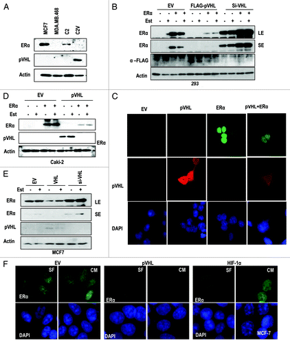
Figure 2. pVHL promotes ER-α degradation through an E3 ligase. (A) Blocking of proteasomal degradation induces ER-α expression. Two hundred and ninety-three cells were transfected with the indicated vectors or si-RNAs for 24 h and were treated with ALLN and MG132 (50 μg/ml) for 6 h. SE and LE indicate short exposure and long exposure. (B) Proteasome inhibitors block pVHL-induced reduction in nuclear ER-α. 293 cells were co-transfected with VHL and ER-α for 24 h and incubated with ALLN or MG132 for 6 h. After fixation with Me-OH, 293 cells were stained with anti-ER-α (red), anti-pVHL (green) and DAPI (blue). (C) si-VHL extends the half-life of ER-α. Two hundred and ninety-three cells were co-transfected with ER-α and si-VHL or pVHL for 24 h. To block de novo synthesis, cycloheximide (CHX; 5 μg/ml) was added for the indicated times. Although VHL transfection completely eliminated ER-α, si-VHL extended the ER-α half-life. LE and SE indicate a long exposure and a short exposure, respectively. (D) pVHL can reduce the half-life of ER-α. MCF7 cells were transfected with the indicated vectors or si-RNAs for 24 h. After washing with serum-free medium, de novo synthesis was blocked by cycloheximide treatment for indicated times, and the expression of ER-α was measured. (E and F) ER-α is ubiquitinylated through the E3 ligase activity of pVHL. Immunoprecipitation was performed using HA or ER-α antibodies, and the co-precipitated proteins were analyzed using the indicated antibodies. Whole-cell extracts were obtained from 293 cells transfected with the indicated vectors. (G) ER-α can be suppressed by wild-type but not mutant pVHL. Two hundred and ninety-three cells were transfected with the indicated vectors or si-RNAs for 24 h.
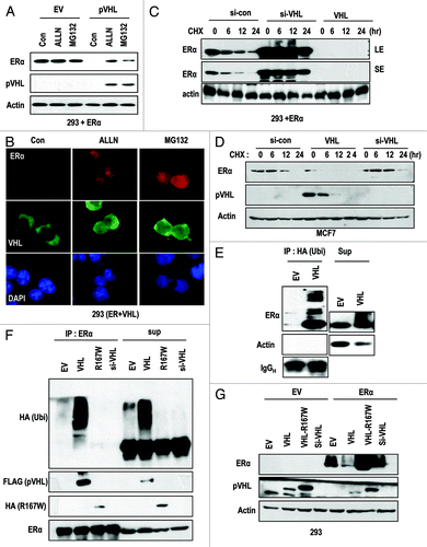
Figure 3. pVHL suppressed ER-α through a direct interaction. (A) Direct interaction between ER-α and pVHL. Immunoprecipitation was performed using a pVHL antibody, and the co-precipitated proteins were analyzed using the indicated antibodies. Whole-cell extracts were obtained from 293 cells transfected with the indicated vectors. (B) GST pull-down assay. Agarose-conjugated GST-pVHL was incubated with whole-cell extracts from 293 cells transfected with the indicated vectors in RIPA buffer for 4 h. PPT indicates proteins that co-precipitated with bead-conjugated pVHL, whereas Sup indicates the supernatant. (C) pVHL suppresses transcription activity of ER-α. Two hundred and ninety-three cells were transfected with ERE-Luc and pVHL or ER-α. The increase in ERE-Luc activity was clearly reduced by VHL transfection. (D) pVHL regulates endogenous ER-α transcription activity. MCF-7 cells were transfected with ERE-Luc and VHL or si-VHL for 24 h. ER-α transcription activity was determined by a luciferase activity. (E) pVHL can suppress cyclin D1 expression. After transfection with the indicated vectors for 24 h, estrogen was added for 6 h in A549 cells. Immunoblot analysis was performed using the indicated antibodies. LE and SE indicate a long exposure and a short exposure, respectively. (F and G) pVHL can suppress Snail expression. After transfection with indicated vectors for 24 h. RT-PCR was performed to determine the expression of Snail using matched specific primers.
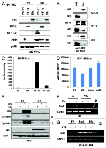
Figure 4. ER-α blocker suppresses cell proliferation in VHL-deficient cells. (A–C) Cell growth was estimated. Each cell line was treated with estrogen or Faslodex (5 μg/ml). Cells were maintained for maximum of 3 d, and cell numbers were calculated daily. (D) VHL-intact cell lines did not respond to estrogen or Faslodex. Each cell line was treated with estrogen or Faslodex for 6 h. After treatment, an immunoblot analysis was performed using the indicated antibodies. (E) The sensitivity to estrogen of Faslodex can be recovered by adding si-VHL to ACHN cells. Cell growth was estimated. After transfection with si-VHL, ACHN cells were treated with estrogen or Faslodex for 6 h. Cells were maintained for 4 d, and cell numbers were calculated daily.
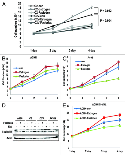
Figure 5. Hypoxia promotes pVHL-mediated ER-α degradation. (A and B) Hypoxia reduces the expression of ER-α in a pVHL-dependent manner. After transfection with the indicated vectors for 24 h, CoCl2 (400 μM) was added for 12 h in each cell line. Immunoblot analysis was performed using the indicated antibodies. (C) pVHL translocates from the cytoplasm to the nucleus under the hypoxic condition. Two hundred and ninety-three cells were transfected with the indicated vectors and were treated with CoCl2 for 12 h. After treatment, cells were harvested to isolate the cytoplasm, membrane/organelle, nucleus and insoluble fractions and these samples were analyzed by immunoblot. (D) Knockdown of ER-α blocks the nuclear translocation of pVHL. Two hundred and ninety-three cells were transfected with the indicated vectors or si-RNAs for 24 h and were treated with CoCl2 for 12 h in 293 cells. After fixation with Me-OH, cells were stained with anti-pVHL (green) and DAPI (blue). (E) ER-α promotes HIF-1α transcription activity. Two hundred and ninety-three cells were co-transfected with an iNOS-Luc reporter containing a HIF-1α response element and the indicated vectors for 24 h. In addition, cells were incubated with CoCl2 for 12 h to induce the hypoxic conditions. (F) Hypoxia-induced suppression of ER-α activity is achieved by pVHL. Two hundred and ninety-three cells transfected with ERE-Luc were incubated with CoCl2 for 12 h. A clear reduction in ERE-Luc activity in response to hypoxia was detected. (G) The resistance of hypoxia-induced ERE-Luc suppression in VHL-deficient cells. Under similar conditions to those described above, a reduction in ERE-Luc activity in response to CoCl2 in C2 cells was not observed. Instead, pVHL transfection could restore sensitivity to hypoxia.
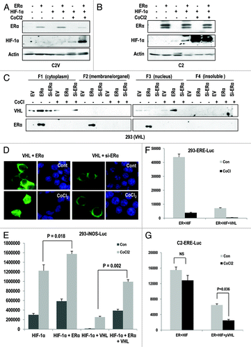
Figure 6. Blocking of ER-α suppresses cell proliferation in VHL-deficient tumors. (A and B) Hypoxia-induced growth arrest is dependent on pVHL status. The growth of cells that lacked VHL (A498; A) was not suppressed by hypoxia. In contrast, ACHN (VHL-positive) cells were sensitive to hypoxia-induced growth suppression. Each cell line was treated with CoCl2. Cells were maintained for 4 d, and cell numbers were calculated daily. (C) The inhibition of ER-α is more effective than hypoxia in cell growth suppression in VHL-deficient cells. C2 cells were incubated in CoCl2 or Faslodex-treated medium for 4 d. (D) Comparison of the growth of C2 and C2V cells. Differentially from C2 cells, where hypoxia did not notably suppress cell growth, C2V cells showed the sensitivity to hypoxia-induced growth suppression. In contrast, the growth of C2 cells was reduced by Faslodex. Cells were incubated with CoCl2 or Faslodex-containing medium for 3 d and fixed with PFA. Cells were visualized by staining with trypan blue. (E and F) pVHL is a critical factor for the determination of hypoxia- or Faslodex-induced growth suppression. Compared with C2V cells, in which Est or Faslodex did not alter cell viability, C2 cells were affected by Faslodex and Est. The opposite effect was observed with CoCl2 treatment. Cells were incubated with the indicated chemicals for 4 d. Cell viability was determined by a MTT assay.
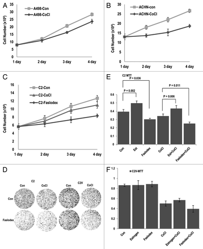
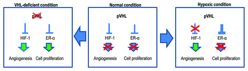
Figure 7. Diagram for the working mechanism. Under normal conditions (center), pVHL suppresses HIF-1α and ER-α through its E3 ligase activity. However, in the hypoxic condition (right panel), pVHL sequesters ER-α more strongly so that cells cannot proliferate. Instead, HIF-1α can be activated and induce angiogenesis. Because the hypoxic condition is generated by tissue damage, this mechanism may contribute to blocking an overgrowth of damaged cells. However, when pVHL is deleted or mutated (left panel), ER-1α-induced cell proliferation can be apparent. In addition, nutrients are then supplied by HIF-1α-mediated angiogenesis. These conditions would increase the probability of the occurrence of cancer.