Figures & data
Figure 1. Inecalcitol induces stronger transcriptional activity than 1,25D3 in SCC cells. (A) SCC or SCC-DR cells were treated with vehicle control EtOH, 1 or 10 nM inecalcitol, or 10 nM 1,25D3 for 48 h. VDR expression was assessed by immunoblot analysis. Actin was the loading control. Results are representative of two independent experiments. (B) 72 h of treatment with inecalcitol induces higher VDR-mediated transcription of the CYP24A1 promoter compared with induction with 1,25D3. Results were normalized to renilla activity as a control for infection. (C–H) SCC cells were treated with EtOH, 1 or 10 nM inecalcitol, or 10 nM 1,25D3 for 24 h unless specified otherwise. mRNA expression of VDR target genes vdr (C), 16 h treatment, p27 (D), 6 h treatment, calbindin D9K (E), amphiregulin (F), p21 (G) and cyclin D1 (H) were assessed by real-time PCR and normalized to mouse gapdh. Results are representative of two independent experiments. *, p < 0.05; **, p < 0.01; ***, p < 0.001.
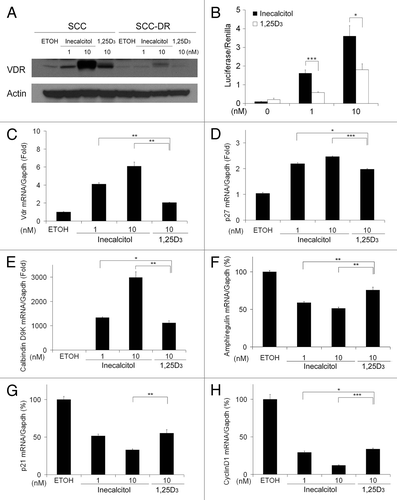
Figure 2. Inecalcitol has greater growth inhibitory effect on SCC than 1,25D3 in vitro. (A) SCC cells were treated with increasing doses of inecalcitol (0.0008–12.5 nM) for 48 h and subjected to MTT assays. Fraction affected (FA) was calculated as 1-(MTT value of the treatment cells)/(MTT value of EtOH-treated cells). (B) Median-effect blot generated using CalcuSyn software to determine the IC50 of inecalcitol. (C) SCC cells were treated with EtOH, inecalcitol, or 1,25D3 and subjected to in vitro clonogenic assaysf. The clones were fixed, stained and counted on day 9. Results are the mean ± SD of triplicate determinations and are representative of three independent experiments. (D and E) SCC (D) or SCC-DR (E) cells were treated with EtOH, 1 or 10 nM inecalcitol, or 10 nM 1,25D3 for 48 h. Cell cycle was examined by PI staining using flow cytometry. Results are representative of three independent experiments.
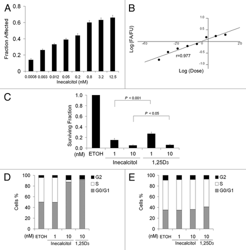
Figure 3. Inecalcitol induces more apoptosis than 1,25D3 in SCC cells. SCC or SCC-DR cells were treated with EtOH, 1 to 100 nM inecalcitol (A) or 1,25D3 (B) for 48 h. Apoptosis was assessed by annexin V-PE and 7AAD staining by flow cytometry. The populations of annexin V−/7AAD−, annexin V+/7AAD− and annexin V+/7AAD+ correspond to live cells, early apoptotic cells and late apoptotic cells, respectively. Representative histograms are presented. The results were summarized in bar graphs as mean ± s.e.m. of annexin V+/7AAD- cells or annexin V+ cells in a representative experiment. *, p < 0.05; **, p < 0.01; ***, p < 0.001. Treatment vs. control “0.”
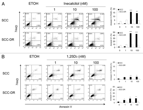
Figure 4. Inecalcitol induces the activation of caspases 8/10 and 3. SCC or SCC-DR cells were treated with EtOH, 1 or 10 nM inecalcitol, or 10 nM 1,25D3 for 48 h. (A) The levels of caspases 8, 10, 9, 3 and PARP were evaluated by immunoblot analysis. Actin was the loading control. (B–E) The activities of caspase 8 (B), 10 (C), 9 (D) and 3 (E) were examined using substrate-based caspase activity assays. Results are representative of three independent experiments. *, p < 0.05; **, p < 0.01.
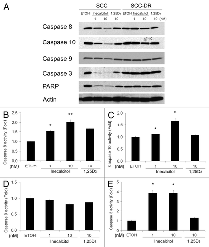
Figure 5. Inecalcitol reduces the expression of c-IAP1 and XIAP. SCC or SCC-DR cells were treated with EtOH, 1 or 10 nM inecalcitol, or 10 nM 1,25D3 for 48 h (A and B) or 24 h (C and D). (A) The protein levels of Bcl-2, Bcl- XL, Mcl-1 and Bax were evaluated by immunoblot analysis. Actin was the loading control. (B) c-IAP1, c-IAP2 and XIAP levels were evaluated by immunoblot analysis. Actin was the loading control. (C and D) mRNA expression levels of c-IAP1 (C) and XIAP (D) were assessed by qRT-PCR and the value is normalized to the mRNA level of Gapdh. Results are representative of three independent experiments. *, p < 0.05; **, p < 0.01; ***, p < 0.001. Treatment vs. EtOH: #, p < 0.05; ##; p < 0.01.
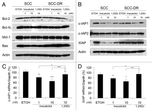
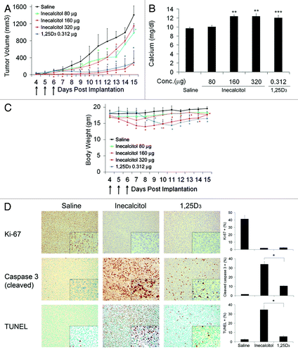
Figure 6. In vivo antitumor activity of inecalcitol and 1,25D3. Four days post inoculation, SCC tumor-bearing C3H mice were treated with saline, 80, 160 or 320 μg/mouse inecalcitol or 0.312 μg/mouse 1,25D3 daily for 3 d. (A) Tumor growth was monitored and measurements taken on the days indicated. Arrows indicate the days animals received treatment. Tumor volumes were calculated by the following formula: volume = (length × width2)/2. (B) Blood was collected 24 h after the last treatment, and serum calcium levels were detected using a colorimetric calcium assay kit. (C) Body weights of the mice were monitored on the days indicated. Arrows indicate the days animals received treatment. (D) SCC tumors in mice treated with 80 μg inecalcitol or 0.312 μg 1,25D3 were harvested at the end of the study and tissues stained with antibodies for Ki-67 and cleaved caspase 3 for immunohistochemistry study or subjected to TUNEL assay. The percentage of positive stained cells were quantified and presented in bar graphs next to the representative images. (×200 and ×400) *, p < 0 0.05; **, p < 0.01; ***, p < 0.001, Treatment vs. saline.