Figures & data
Figure 1. HU stimulates the interaction between PLK1 and BRCA1. (A–C) U2-OS cells were either left untreated (−) or treated with 4 mM HU for 24 h (+). Equal amount of cell lysate was used for immunoprecipitation (IP). The antibodies used for IP are indicated on the top. PLK1m is a mouse monoclonal antibody. PLKR is a rabbit polyclonal antibody. IgG was used as the negative control. Immunoblotting (IB) antibodies are indicated on the right. (D) Depletion of BRCA1 by siRNA abolishes the interaction between PLK1 and BRCA1. U2-OS cells were first transfected with either control siRNA, or two different siRNA against BRCA1 (BRCA1-B or BRCA1-C), and then treated with 4 mM HU for 24 h. Equal amount of cell lysate was used for IP. IB antibodies are indicated on the right. (E) The PLK1-BRCA1 interaction is independent of the kinase activity of PLK1 and Aurora A. U2-OS cells were first treated with 4 mM HU for 24 h. 100 nM BI2536 or 3 μM MLN8054 was added to the medium, and cells were incubated for another 24 h. Equal amount of cell lysate was used for IP. IB antibodies are indicated on the right. (F) GFP-tagged BRCA1 and Myc-tagged hBora were con-transfected into 293T cells. Cell lysates were used for IP with either control IgG or anti-Myc antibody. Antibodies used for immunoblotting are indicated on the right.
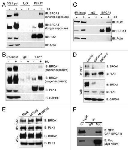
Figure 2. PLK1 interacts with both the RING domain and the BRCT domain of BRCA1, while hBora primarily binds the RING domain of BRCA1. (A) Diagram of the domain structure of BRCA1 and the summary of the domain mapping. (B) HA-PLK1 and GFP-tagged full-length BRCA1 or different variants were co-transfected into 293T cells. Cell lysates were used for IP with anti-HA antibody. IB antibodies are indicated on the right. (C) Myc-hBora and GFP-tagged full-length BRCA1 or different variants were co-transfected into 293T cells. Cell lysates were used for IP with anti-Myc antibody. IB antibodies are indicated on the right. WCL, whole-cell lysate.
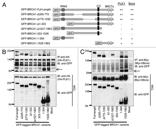
Figure 3. HU inhibits the kinase activity of PLK1 and induces dynamic interactions of PLK1, hBora, and BRCA1. U2-OS cells were either left untreated (0 h) or treated with 4 mM HU for the indicated time. Equal amount of cell lysate was used for IP with antibody against either PLK1 (A and B) or phosphorylated T-210 of PLK1 (D and E) (pT210-PLK1). IB antibodies are indicated on the right. (C) U2-OS cells were either left untreated (0 h) or treated with 4 mM HU for the indicated time. Equal amount of cell lysate was incubated with either λ phosphatase or phosphatase inhibitor at 30 °C for 30 min. Cell lysates were then run on a SDS-PAGE and immunoblotted with antibody against hBora. WCL, whole-cell lysate.
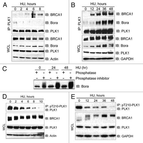
Figure 5. Depletion of BRCA1 impairs the inhibition of PLK1 activity in HU treated cells. (A) Depletion of BRCA1 induces hyper-phosphorylation of hBora. U2-OS cells were first transfected with either control siRNA, or one of two different siRNAs against BRCA1 (BRCA1-B or BRCA1-C). Cells were then either left untreated (0 h) or treated with 4 mM HU for the indicated times. Equal amount of cell lysate was then run on a SDS-PAGE and immunoblotted with the antibody indicated on the right. (B) U2-OS cells were first transfected with either control siRNA, or one of two different siRNAs against BRCA1 (BRCA1-B or BRCA1-C). Cells were either left untreated or treated with 4 mM HU for 48 h. Equal amount of cell lysate was used for IP with antibody against pT210-PLK1. IB antibodies are indicated on the right. (C) U2-OS cells were first transfected with either control siRNA, or one of two different siRNAs against BRCA1 (BRCA1-B or BRCA1-C). Cells were then treated with 4 mM HU for 48 h. Equal amount of cell lysate was used for IP with either mouse IgG or an antibody against PLK1 (left panel). The immunoprecipitated PLK1 was then used for kinase assay using GST-PBIPtide as the substrate and in the presence of [γ-32P]-ATP (right panel). Samples were then run on a 4–12% SDS-PAGE. Phosphorylated GST-PBIPtide was visualized by autoradiography. The amount of PLK1 that had been pulled down was determined by immunoblotting with an antibody against PLK1. The intensity of the 32P-GST-PBIPtide was quantified on a Typhoon Phosphoimager and ImageQuant software.
![Figure 5. Depletion of BRCA1 impairs the inhibition of PLK1 activity in HU treated cells. (A) Depletion of BRCA1 induces hyper-phosphorylation of hBora. U2-OS cells were first transfected with either control siRNA, or one of two different siRNAs against BRCA1 (BRCA1-B or BRCA1-C). Cells were then either left untreated (0 h) or treated with 4 mM HU for the indicated times. Equal amount of cell lysate was then run on a SDS-PAGE and immunoblotted with the antibody indicated on the right. (B) U2-OS cells were first transfected with either control siRNA, or one of two different siRNAs against BRCA1 (BRCA1-B or BRCA1-C). Cells were either left untreated or treated with 4 mM HU for 48 h. Equal amount of cell lysate was used for IP with antibody against pT210-PLK1. IB antibodies are indicated on the right. (C) U2-OS cells were first transfected with either control siRNA, or one of two different siRNAs against BRCA1 (BRCA1-B or BRCA1-C). Cells were then treated with 4 mM HU for 48 h. Equal amount of cell lysate was used for IP with either mouse IgG or an antibody against PLK1 (left panel). The immunoprecipitated PLK1 was then used for kinase assay using GST-PBIPtide as the substrate and in the presence of [γ-32P]-ATP (right panel). Samples were then run on a 4–12% SDS-PAGE. Phosphorylated GST-PBIPtide was visualized by autoradiography. The amount of PLK1 that had been pulled down was determined by immunoblotting with an antibody against PLK1. The intensity of the 32P-GST-PBIPtide was quantified on a Typhoon Phosphoimager and ImageQuant software.](/cms/asset/aad7ddfe-1f65-4fec-9bc6-483853d34413/kccy_a_10925349_f0004.gif)
Figure 4. Genotoxic stresses induce strong interaction between PLK1 and BRCA1. (A) Two sets of U2-OS cells were treated with 4 mM HU. Twelve hours later, 5 mM caffeine was added to one set of cells. Twelve hours after this addition, equal amount of cell lysate was used for IP with antibody against PLK1. IB antibodies are indicated on the right. Arrows indicate the IgG heavy chain. (B and C) U2-OS cells were either treated with vehicle (mock), or with 4 mM HU, 3 μM aphidicolin, 50 μM etoposide, 1 μM Daunorubicin, or 5 μM cis-platin (CDDP) for 24 h. Equal amount of cell lysate was used for IP with antibody against either PLK1 (B) or pT210-PLK1 (C). IB antibodies are indicated on the right. WCL, whole-cell lysate.
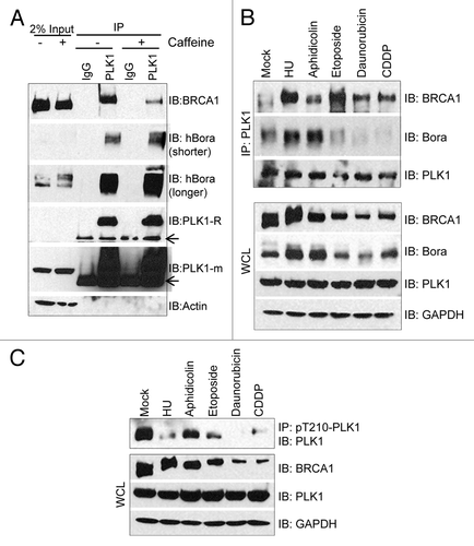
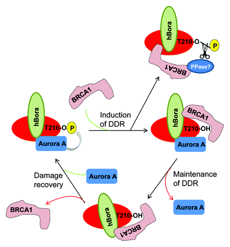
Figure 6. A Model of how BRCA1 inhibits PLK1 activity during DDR.