Figures & data
Figure 1. Acute treatment with rapamycin alters polysome profile in mouse liver tissue. (A) Example liver polysome profile. Polysome gradients contain peaks representing an insoluble fraction, free ribosome subunits (40s and 60S), and active ribosomes (R) separated by the amount of ribosomes tethered to mRNA. (B) Acute rapamycin treatment involved a single injection of vehicle or rapamycin (8 mg/kg). Tissue was harvested 1 h after injection. (C and D) representative liver polysome profiles from vehicle (C) and rapamycin (D) treated mice. Arrows indicate 40 and 60S peaks. (E) Quantification of polysome peaks from vehicle and rapamycin-treated liver tissue. *P < 0.05; 2-way ANOVA, Bonferroni post hoc test. For both groups, n = 8.
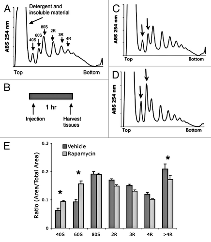
Figure 2. Acute treatment with rapamycin alters polysome profile in mouse muscle tissue. (A) Example muscle polysome profile. (B and C) representative muscle polysome profiles from vehicle (B) and rapamycin (C) treated mice. Arrows indicate 60S and 80S peaks. (D) Quantification of polysome peaks from vehicle and rapamycin-treated liver tissue. ***P < 0.001; *P < 0.05; 2-way ANOVA, Bonferroni post hoc test. For both groups, n = 6.
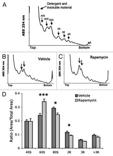
Figure 3. ARA demonstrates that acute rapamycin reduces ribosome activity. (A) Example liver ARA profile. Unlike polysome gradients, active ribosomes are combined into a single peak. (B) Quantification of liver ARA from vehicle and rapamycin-treated mice. (C) Example muscle ARA profile. (D) Quantification of muscle ARA from vehicle and rapamycin-treated mice. ***P < 0.001; **P < 0.05; 2-way ANOVA, Bonferroni post hoc test. For all groups, n = 6.
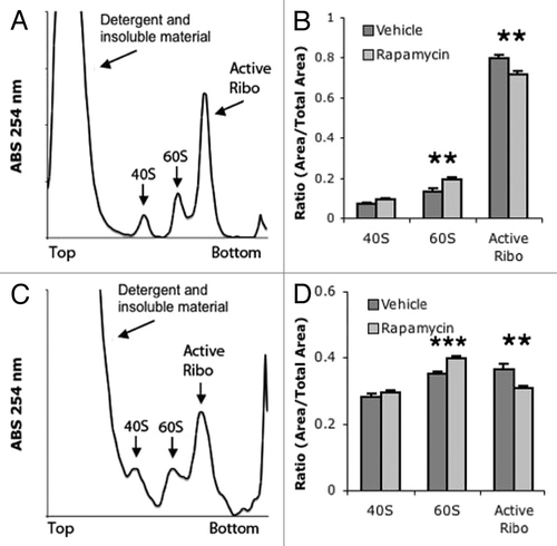
Figure 4. Chronic treatment with rapamycin does not alter polysome profiles in mouse tissue. (A) Chronic treatment involved an injection every 48 h with vehicle or rapamycin. Tissue was collected 1 h after the final injection. (B and C) representative liver polysome profiles from vehicle (B) and rapamycin (C) treated mice. (D) Quantification of polysome peaks from vehicle and rapamycin-treated liver tissue. (E and F) representative muscle polysome profiles from vehicle (E) and rapamycin (F) treated mice. (G) Quantification of polysome peaks from vehicle and rapamycin-treated liver tissue. For all groups, n = 8.
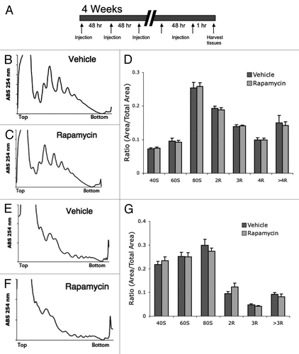
Figure 5. No difference in ARA profiles between chronic vehicle and rapamycin-injected mice. (A) Quantification of liver ARA from chronically vehicle and rapamycin-treated mice. (B) Quantification of muscle ARA from chronically treated vehicle and rapamycin-treated mice. For all groups, n = 6.
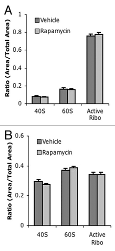
Figure 6. Acute and chronic rapamycin treatments reduce levels of phospho S6K1 and phospho S6. (A– C) Western blot analysis of liver following acute treatment with vehicle or rapamycin. (A) Representative blots showing westerns against phospho-S6K1 (T389) and total S6K1 and phospho-S6 (S240/S244) in vehicle (Veh) or rapamycin (Rap) treated liver. (B) Quantification of phospho-S6K1 to total S6K1. (C) Quantification of phospho-S6 to total S6. (D–F) western blot analysis of muscle following acute treatment with vehicle or rapamycin. (D) Representative blots (E) Quantification of phospho S6K1 to total S6K1. (F) Quantification of phospho-S6 to total S6. (G–I) Western analysis of liver following chronic treatment with vehicle or rapamycin. (G) Representative blots. (H) Quantification of phospho S6K1 to total S6K1. (I) Quantification of phospho-S6 to total S6. (J–L) Western analysis of muscle following chronic treatment with vehicle or rapamycin. (G) Representative blots. (E) Quantification of phospho S6K1 to total S6K1. (F) Quantification of phospho-S6 to total S6. T-test was used to compare Veh to Rap: *P < 0.05; **P < 0.01; ***P < 0.001. For all experiments n = 4 samples per group.
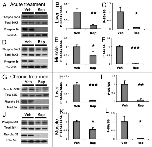
Figure 7. Acute and chronic rapamycin treatments increase levels of hypo-phosphorylated 4EBP. (A) western blots of 4EBP show 3 bands, hyperphosphorylated high weight (gamma) and middle (beta) bands, and a lower weight hypo-phosphorylated (alpha) band. (B–D) Western blot analysis of liver and muscle following acute treatment with vehicle or rapamycin. (B) Representative western blots of liver and muscle tissues following acute vehicle (Veh) or rapamycin (Rap) treatment. (C) Quantification of the ratio of hypo-phosphorylated alpha band of 4EBP over the total 4EBP bands in liver tissue. (D) Quantification of the ratio of alpha to total 4EBP bands in muscle tissue. (E–G) western blot analysis of liver and muscle following chronic treatment with vehicle or rapamycin. (E) Representative western blots of liver and muscle tissues following chronic vehicle or rapamycin treatment. (F) Quantification of the ratio of alpha to total 4EBP bands in liver tissue following chronic treatments. (G) Quantification of the ratio of alpha to total 4EBP bands in liver tissue following chronic treatments. T-test was used to compare Veh to Rap: *P < 0.05; **P < 0.01; ***P < 0.001. For all acute treated groups, n = 8. For all chronic vehicle groups, n = 8. For chronic rapamycin groups, n = 6.
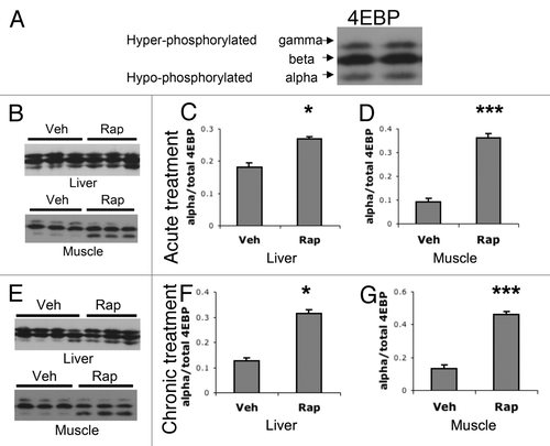
Figure 8.S6K1−/− mice do not have altered polysome profiles. (A and B) representitive liver polysome profiles from S6K1+/+ mice (A) and S6K1−/− mice (B). (C) Quantification of polysome peaks from S6K1+/ + mice and S6K1−/− liver tissue. For both groups, n = 10. (D and E) representative muscle polysome profiles from S6K1+/+ mice (D) and S6K1−/− mice (E). (F) Quantification of polysome peaks from S6K1+/+ mice and S6K1−/− liver tissue. (G and H) No difference in ARA profiles between chronic vehicle and rapamycin-injected mice. (G) Quantification of liver ARA from S6K1+/+ mice and S6K1−/− mice. (H) Quantification of muscle ARA from S6K1+/+ mice and S6K1−/− mice. For all groups, n = 6.
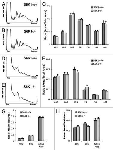
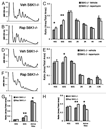
Figure 9. Acute rapamycin treatment reduces translation in S6K1−/− mice. (A and B) representative liver polysome profiles from S6K1+/+ mice (A) and S6K1−/− mice (B). (C) Quantification of polysome peaks from S6K1+/+ mice and S6K1−/− liver tissue, vehicle n = 6, rapamycin n = 5. (D and E) representative muscle polysome profiles from S6K1+/+ mice (D) and S6K1−/− mice (E). (F) Quantification of polysome peaks from S6K1+/+ mice and S6K1−/− liver tissue, for both groups n = 6.. (G and H) ARA profiles between chronic vehicle and rapamycin-injected mice. (G) Quantification of liver ARA from S6K1+/+ mice and S6K1−/− mice. (H) Quantification of muscle ARA from S6K1+/+ mice and S6K1−/− mice. For all groups, n = 6. **P < 0.01; *P < 0.05; 2-way ANOVA, Bonferroni post hoc test.