2,760
Views
43
CrossRef citations to date
0
Altmetric
Report

Structure-based discovery of the first allosteric inhibitors of cyclin-dependent kinase 2

, , , &
Pages 2296-2305 | Received 23 Dec 2013, Accepted 20 May 2014, Published online: 09 Jun 2014

Figures & data

Chart 1. Chemical structures of the allosteric inhibitors 1–7 and those resulting from the hit expansion of compound 4.
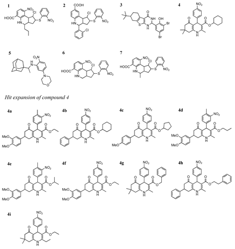
Chart 1. Chemical structures of the allosteric inhibitors 1–7 and those resulting from the hit expansion of compound 4.

Table 1. Predicted free energies of binding, VS rankings, chemical classification, occupation of the ANS1 and ANS2 pockets, selected physico–chemical properties, and biological properties of the 7 allosteric inhibitors discovered in the primary virtual screening (compounds 1-7) and of the nine analogs of compound 4 selected from hit expansion (compounds 4a-4i)

Figure 1. Concentration-dependent displacement of ANS from CDK2. Panel (A) reports the displacement activity of compounds 1–7, while (B) shows the activity of six (4b–4g) of the nine compounds derived from the hit expansion of compound 4. Compounds 4a, 4h, and 4i did not show appreciable displacement activity.
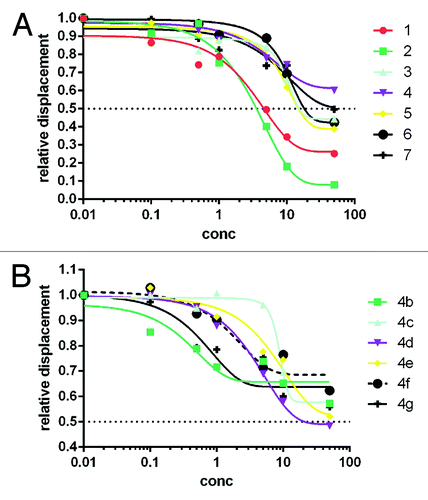
Figure 1. Concentration-dependent displacement of ANS from CDK2. Panel (A) reports the displacement activity of compounds 1–7, while (B) shows the activity of six (4b–4g) of the nine compounds derived from the hit expansion of compound 4. Compounds 4a, 4h, and 4i did not show appreciable displacement activity.

Figure 2. Inhibition of Rb phosphorylation by compound 4 and SU9516 in MDA-MB231 cells. Proteins were separated on SDS page, electroblotted to nitrocellulose, and hybridized with antibodies against phoshorylated Rb (T821, left panel and S780, right panel). Blots were subsequently hybridized with antibodies against total Rb and actin (used as loading control). C, control, untreated cells; SU, cells treated with SU9516 (10 μM); 4, cells treated with compound 4 (4 μM).
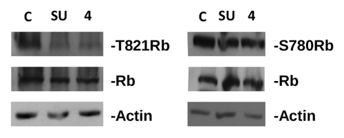
Figure 2. Inhibition of Rb phosphorylation by compound 4 and SU9516 in MDA-MB231 cells. Proteins were separated on SDS page, electroblotted to nitrocellulose, and hybridized with antibodies against phoshorylated Rb (T821, left panel and S780, right panel). Blots were subsequently hybridized with antibodies against total Rb and actin (used as loading control). C, control, untreated cells; SU, cells treated with SU9516 (10 μM); 4, cells treated with compound 4 (4 μM).

Figure 3. Predicted binding modes of compounds 1–7 in the allosteric pocket of CDK2, highlighting (left) the location of each compound in the overall fold, (right) the close up of the allosteric site with selected residues interacting with the ligands. For comparative purposes, the first box shows the binding mode of the 2 ANS molecules present in the crystal structure of the binary complex (PDB code 1PXF).
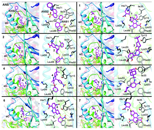
Figure 3. Predicted binding modes of compounds 1–7 in the allosteric pocket of CDK2, highlighting (left) the location of each compound in the overall fold, (right) the close up of the allosteric site with selected residues interacting with the ligands. For comparative purposes, the first box shows the binding mode of the 2 ANS molecules present in the crystal structure of the binary complex (PDB code 1PXF).
Supplemental material

Additional material

Download Zip (1.2 MB)

Reprints and Corporate Permissions

Please note: Selecting permissions does not provide access to the full text of the article, please see our help page How do I view content?

To request a reprint or corporate permissions for this article, please click on the relevant link below:

Academic Permissions

Please note: Selecting permissions does not provide access to the full text of the article, please see our help page How do I view content?

Obtain permissions instantly via Rightslink by clicking on the button below:

If you are unable to obtain permissions via Rightslink, please complete and submit this Permissions form. For more information, please visit our Permissions help page.