Figures & data
Figure 1 All roads lead to JNK. Diagram with the proposed mechanisms for the activation of JNK after the loss of scrib group genes. JNK pathway activation normally results in the death of the mutant cells; however, in the presence of oncogenic Ras JNK can promote tumor progression. See the text for details.
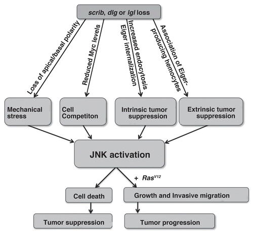
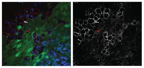
Figure 2 Eiger/dTNF is primarily expressed by tumor-associated hemocytes. The confocal image displays a RasV12/scrib imaginal disc tumor. The tumor cells are labelled with GFP (green), Eiger immunostaining is in red and nuclei are shown in blue. The Eiger staining is shown in grey in the lower part. The white arrow points to a tumor-associated cell that expresses high levels of Eiger. These tumor-associated cells were identified as plasmatocytes, a sub-type of haemocytes.Citation23 The red arrow points to a tumor cell displaying Eiger staining in the form of intracellular puncta, identified previously as endocytic compartments.Citation22