Figures & data
Figure 1. Identification of epiplexus cells as immune cells. (A) Scheme of the experimental approach. (B) CPs are located in all four brain ventricles. (C) An acutely isolated and intact CP from the lateral ventricle. Scale bar is approximately 1 mm. (D) A living epiplexus cell resting on the choroidal epithelium, visualized with transmitted light (left) or by labeling with Alexa Fluor 488 isolectin B4 conjugate from Griffonia simplicifolia (center); EC – epiplexus cell, EP – choroidal epithelium, BV – blood vessel, LV – lumen of the lateral ventricle. Scale bar is 20 μm. (E) Immunofluorescent staining for immune cell markers, Iba1 and IB4 in the CP. Note the significant co-localization (yellow cells in the merge image). Scale bar is 20 μm.
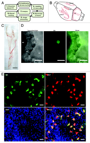
Figure 2. Extracellular ATP triggers chemokinesis of epiplexus cells. (A and D) Representation of the tracked paths superimposed on the original image under control conditions and in the presence of exogenous 100 µM ATP. Labels in the top right of the images represent the time relative to the start of the experiment (0 min). Note the 25 min was the end of the baseline and 120 min was the end of the experiment. Scale bar is 50 µm. Raw data showing the distance traveled by individual epiplexus cells in control (B) and in the presence of ATP (E). Each colored line represents an individual epiplexus cell. (B and E) Raw distance traveled during 5 min intervals; (C and F) normalized summative distance traveled.
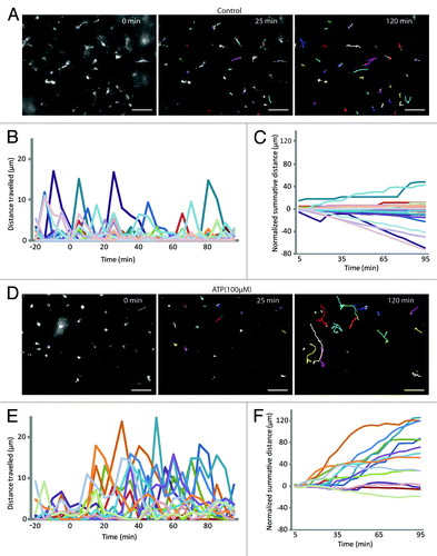
Figure 3. Panx1 channels are involved in epiplexus cells activation by exogenous 100 µM ATP. Panx1 channels are robustly expressed on choroidal epithelium, but were rarely detected in epiplexus cells. Western blotting analysis confirmed presence of Panx1 in the CP. The Panx1 blockers, 500 µM probenecid and 100 µM 10panx significantly decreased ATP-triggered chemokinesis. (A and B) Immunofluorescent staining for Panx1 in the CP and on an individual IB4-positive epiplexus cell. (C) Detection of Panx1 protein by western blotting. (D) Normalized average distance and statistical analysis, each symbol represents a single cell and all cells from the experiments are shown; (E) normalized summative distance; (F) slope of normalized summative distance where each symbol represents a single isolated CP. Scale bars are 50 μm (A) and 5 μm (B).
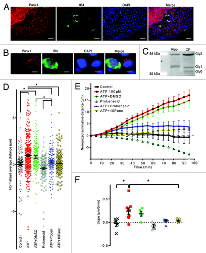
Figure 4. P2X7 and P2X4 receptors are both expressed on epiplexus cells, but only P2X4 is involved in chemokinesis. (A) Immunofluorescent staining for P2X4 receptors in epiplexus cells and CP epithelium. Note that both the epithelial and the epiplexus cells are positive for P2X4. (B) Immunofluorescent staining for P2X7 receptors is evident only in epiplexus cells. (C, D, and E) The P2X7 receptor blocker 1 µM BBG did not prevent activation of epiplexus cell by ATP, and the P2X7 receptor agonist BzATP did not trigger an increase in epiplexus cell activity. Thirty µM 5-BDBD, a P2X4 receptor antagonist, alone or in combination with probenecid significantly decreased ATP-triggered chemokinesis. (C) Normalized average distance and statistical analysis where each symbol represents an individual cell; (D) normalized summative distance; (E) slope of normalized summative distance where each symbol represents the average of all cells in a given CP. Scale bars are 50 μm.
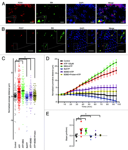
Figure 5. Ca2+ is involved in mediating chemokinesis. Absence of extracellular Ca2+ and chelation of intracellular Ca2+ significantly decreased ATP-triggered chemokinesis. (A) Normalized average distance and statistical analysis, each symbol represents an individual cell; (B) normalized summative distance; (C) slope of normalized summative distance where each symbol represents the average of all cells in a given CP.
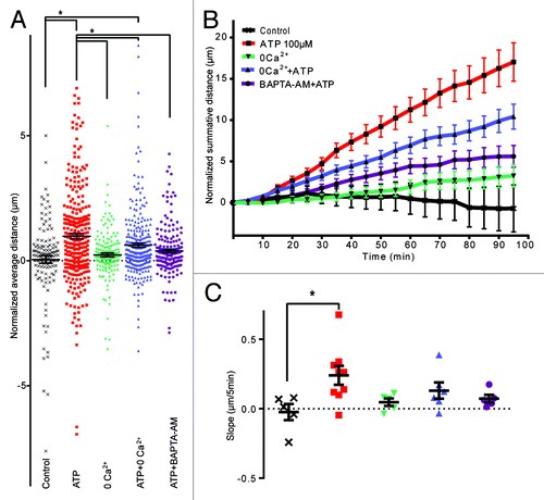
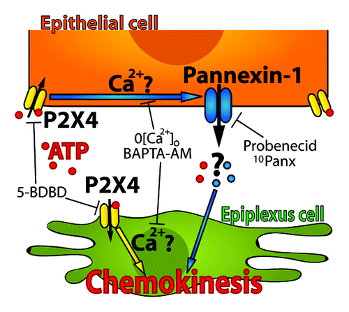
Figure 6. A model of a proposed cascade for ATP-triggered chemokinesis of epiplexus cells. P2X4 receptors on the epithelial cells may be functionally coupled to Panx1 channels. In this way, activation of P2X4 receptors by ATP would cause a release of an unidentified signaling molecule though Panx1 channels to activate chemokinesis of epiplexus cells. Our current data cannot exclude the possibility that P2X4 receptors on the epiplexus cells may contribute to the activation process.