Figures & data
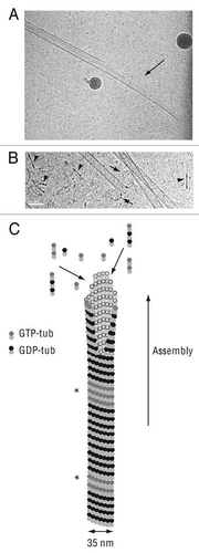
Figure 1 Microtubule assembly. (A) Cryo-electron microscopy image of a growing microtubule end showing an opened sheet (arrow). (B) Cryoelectron microscopy image of a shrinking microtubule end showing protofilaments peeling into ring-like structures (arrows). Numerous tubulin oligomers coming from microtubule disassembly are visible in the background (arrowheads). Scale bar, 50 nm in both parts. (C) Schematic representation of a growing microtubule. The end displays a sheet-like conformation which later close into a tube. GTP-tub dimers, GDP-dimers and tubulin oligomers participate to microtubule elongation at the end of the polymer. GTP-tub, α-tubulin and β-tubulin associated to GTP; GDP-tub, α-tubulin and β-tubulin associated to GDP; *indicates GTP-remnants integrated in the microtubule wall. Cryo-electron microscopy images are courtesy of Dr. I. Arnal, UMR 6026, Rennes, France.