297
Views
4
CrossRef citations to date
0
Altmetric
Article Addendum

A question of homology for chordate adhesive organs

&
Pages 75-77 | Received 12 Oct 2010, Accepted 12 Oct 2010, Published online: 01 Jan 2011

Figures & data

Figure 1 Comparison of the Xenopus cement gland and the Astyanax casquette. Basic properties of the two organs (black) are compared: position, gene expression, trigeminal innervation and functional role. Only the former is different between the two species.
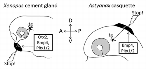
Figure 1 Comparison of the Xenopus cement gland and the Astyanax casquette. Basic properties of the two organs (black) are compared: position, gene expression, trigeminal innervation and functional role. Only the former is different between the two species.

Figure 2 Adhesive organs in the chordate phylogenetic tree. A simplified chordate tree helps visualisation of the presence of adhesive organs in diverse taxa, as well as the extreme diversity of their morphology. Position, number, size, shape and structure vary greatly between examples shown. A star indicates that adhesive organs have also been described in these species; however they are not illustrated for the sake of clarity of the figure. Drawing credits: the Lepidosteus (gar) sucking disk is a detail taken from Alexander Agassiz, Planche II.Citation11 The Xenopus laevis cement gland is a detail from the stage 38 embryo of the Nieuwkoop and Faber staging table.Citation18 The salamander (Pleurodeles Waltl) balancer is a detail taken from Figure 5.12 of Duellman and Trueb.Citation19 The ascidian papilla, the Astyanax casquette and the Tilapia adhesive glands were drawn by SR. A blind cavefish is drawn for Astyanax, so that the eye is small and transparent, hence not visible.
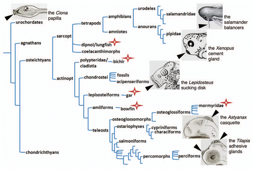
Figure 2 Adhesive organs in the chordate phylogenetic tree. A simplified chordate tree helps visualisation of the presence of adhesive organs in diverse taxa, as well as the extreme diversity of their morphology. Position, number, size, shape and structure vary greatly between examples shown. A star indicates that adhesive organs have also been described in these species; however they are not illustrated for the sake of clarity of the figure. Drawing credits: the Lepidosteus (gar) sucking disk is a detail taken from Alexander Agassiz, Planche II.Citation11 The Xenopus laevis cement gland is a detail from the stage 38 embryo of the Nieuwkoop and Faber staging table.Citation18 The salamander (Pleurodeles Waltl) balancer is a detail taken from Figure 5.12 of Duellman and Trueb.Citation19 The ascidian papilla, the Astyanax casquette and the Tilapia adhesive glands were drawn by SR. A blind cavefish is drawn for Astyanax, so that the eye is small and transparent, hence not visible.