517
Views
13
CrossRef citations to date
0
Altmetric
Article Addendum

Differential localization of SAP102 and PSD-95 is revealed in hippocampal spines using super-resolution light microscopy

, , &
Pages 104-105 | Received 08 Nov 2010, Accepted 10 Nov 2010, Published online: 01 Jan 2011

Figures & data

Figure 1 Distribution of SAP102 and PSD-95 in spines. (A–C) Double labeling of endogenous SAP102 and PSD-95 in hippocampal neurons (21 DIV). (A) Image was taken with a conventional confocal microscope. The spine in the upper left is enlarged and shown in the bottom left. The asterisk indicates the dendrite region and the arrows indicate spines. Scale bars, 500 nm. (B) Images were taken with a Leica STED microscope. For the spines in the top and the middle parts, PSD-95 (red) was imaged in the STED channel and SAP102 (green) was imaged in the regular channel. For the spine in the bottom part, SAP102 (red) was imaged in the STED channel and PSD-95 (green) was imaged in the regular channel. Scale bars, 500 nm. (C) Images were taken with a DeltaVision 3D super resolution microscope. The top, middle and bottom parts are images of the same spine viewed in 3D at three different angles. The grid squares are 500 nm. (D) Immunogold labeling of SAP102 (5 nm gold) and PSD-95 (15 nm gold) in synapses from cultured hippocampal neurons (21 DIV). SAP102 is distributed in both the PSD (arrowhead) and cytoplasm (arrow), while PSD-95 is concentrated in the PSD area. P, presynaptic terminal. Scale bar, 100 nm.
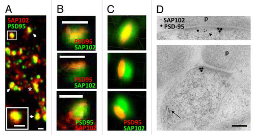
Figure 1 Distribution of SAP102 and PSD-95 in spines. (A–C) Double labeling of endogenous SAP102 and PSD-95 in hippocampal neurons (21 DIV). (A) Image was taken with a conventional confocal microscope. The spine in the upper left is enlarged and shown in the bottom left. The asterisk indicates the dendrite region and the arrows indicate spines. Scale bars, 500 nm. (B) Images were taken with a Leica STED microscope. For the spines in the top and the middle parts, PSD-95 (red) was imaged in the STED channel and SAP102 (green) was imaged in the regular channel. For the spine in the bottom part, SAP102 (red) was imaged in the STED channel and PSD-95 (green) was imaged in the regular channel. Scale bars, 500 nm. (C) Images were taken with a DeltaVision 3D super resolution microscope. The top, middle and bottom parts are images of the same spine viewed in 3D at three different angles. The grid squares are 500 nm. (D) Immunogold labeling of SAP102 (5 nm gold) and PSD-95 (15 nm gold) in synapses from cultured hippocampal neurons (21 DIV). SAP102 is distributed in both the PSD (arrowhead) and cytoplasm (arrow), while PSD-95 is concentrated in the PSD area. P, presynaptic terminal. Scale bar, 100 nm.