Figures & data
Figure 1 Cartoon diagrams of genetically encoded force sensors where the letter A in the fluorophore represents the acceptor Venus, the letter D represents the donor Cerulean. The arrows represent energy transfer from donor to acceptor. (A) stFRET; (B) sstFRET. (C) TSMod, mTFP1 is the donor and Venus is the acceptor. (D) PriSSM, using wild-type GFP and circularly permutated GFP for PRIM dimers.
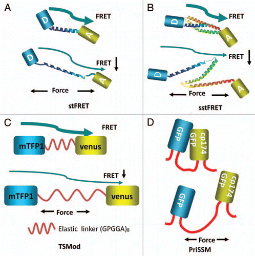
Figure 2 Diagrams of inserting the force probes into host proteins. (A) Actinin-stFRET forming anti-parallel dimers. (B) Spectrin-stFRET exists as a hetero-dimer with α and β subunits. (C) Vinculin-TSMod associating at focal adhesions as monomers. (D) A protein construct of PriSSM with myosin motors on the ends.
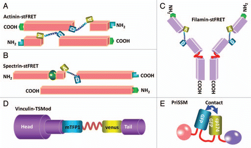
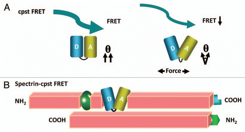
Figure 3 A new and smaller FRET force sensor (cpstFRET) with higher sensitivity and wider dynamic range. (A) shows applied force twisting the fluorophores out of alignment and lower FRET. (B) shows cpstFRET incorporated into a spectrin α subunit that is part of a heterodimer.