Figures & data
Figure 1 Glutathione depletion is associated with 3-nitrotyrosine increase in mice brain without accumulation of apoptotic markers. (A) Left part: 20 µg of proteins extracted from mice brains were spotted on nitrocellulose membrane and subjected to Dot blot analysis using a polyclonal 3-nitrotyrosine (3-NT) antibody. Right part: density of immunoreactive dots was normalized for Ponceau Red spots and reported as arbitrary units (a.u.) and as means ± SD. (*p < 0.01 versus control, **p < 0.01 versus BSO-treated; n = 3). (B) 20 µg of total protein extracts were loaded for detection of Bcl-2, Bax and pro-caspase-3 by western blot. Actin was used as loading control. (C) mitochondria were purified from mice brains homogenates by Percoll® gradient, lysed and 20 µg of proteins were loaded for detection of AIF by western blot. Cytochrome c was used as loading control.
![Figure 1 Glutathione depletion is associated with 3-nitrotyrosine increase in mice brain without accumulation of apoptotic markers. (A) Left part: 20 µg of proteins extracted from mice brains were spotted on nitrocellulose membrane and subjected to Dot blot analysis using a polyclonal 3-nitrotyrosine (3-NT) antibody. Right part: density of immunoreactive dots was normalized for Ponceau Red spots and reported as arbitrary units (a.u.) and as means ± SD. (*p < 0.01 versus control, **p < 0.01 versus BSO-treated; n = 3). (B) 20 µg of total protein extracts were loaded for detection of Bcl-2, Bax and pro-caspase-3 by western blot. Actin was used as loading control. (C) mitochondria were purified from mice brains homogenates by Percoll® gradient, lysed and 20 µg of proteins were loaded for detection of AIF by western blot. Cytochrome c was used as loading control.](/cms/asset/c060cf22-f248-4fc4-8a95-0c1bb2216d7f/kcib_a_10915654_f0001.gif)
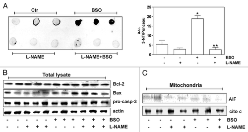
Table 1 GSH concentration in mice treated with BSO