Figures & data
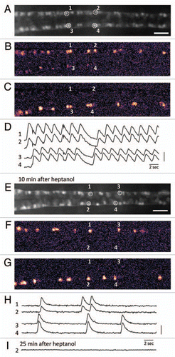
Figure 1 Calcium imaging of the motor circuit in the zebrafish spinal cord during spontaneous contractions. (A) Expression of GCaMP-HS in the CaP motor neurons in the spinal cord. A dorsal view of the SAIG213A;UA S:GCaMPHS double transgenic embryo embedded in 2% low-melting agarose under a fluorescence microscope and treated with a neuromuscular junction blocker, D-tubocurarine. CaP neurons #1–#4 were used as ROIs (regions of interest). Anterior to the left. Scale bar: 200 µm. (B and C) Calcium signals of the CaP motor neurons with pseudocolors (see also www.youtube.com/watch?v=6y44uxrh7z4). (B) The CaP motor neurons on the right side including ROI-1 and -2 showed increased fluorescence. (C) The CaP motor neurons on the left side including ROI-3 and -4 showed increased fluorescence. (D) The fluorescence changes in the selected CaP motor neurons. 1 and 2 (3 and 4) are activated synchronously, and the right (1 and 2) and left (3 and 4) neurons are activated alternately. A vertical bar indicates (F-resting F)/resting F = 50%. (E) A dorsal view of the SAIG213A;UA S:GCaMPHS double transgenic embryo embedded in 2% low-melting agarose under a fluorescence microscope and treated with a gap junction blocker, heptanol, for 10 min. Anterior to the left. Scale bar: 200 µm. (F and G) Calcium imaging of the CaP motor neurons in the presence of heptanol with pseudocolors (see also www.youtube.com/watch?v=3xhw9D35H5w). (F) The CaP motor neurons on the right side including ROI-1 and -2 showed increased fluorescence. (G) The CaP motor neurons on the left side including ROI-3 and -4 showed increased fluorescence. (H) The fluorescence changes in the selected CaP motor neurons. Synchronized activation is still observed. A vertical bar indicates (F-resting F)/resting F = 100%. (I) The fluorescence changes are not detected 25 min after the heptanol treatment.